Welcome to The Visible Embryo

Home-- -History-- -Bibliography- -Pregnancy Timeline- --Prescription Drugs in Pregnancy- -- Pregnancy Calculator- --Female Reproductive System- -Contact
 

Welcome to The Visible Embryo, a comprehensive educational resource on human development from conception to birth.

The Visible Embryo provides visual references for changes in fetal development throughout pregnancy and can be navigated via fetal development or maternal changes.

The National Institutes of Child Health and Human Development awarded Phase I and Phase II Small Business Innovative Research Grants to develop The Visible Embryo. Initally designed to evaluate the internet as a teaching tool for first year medical students, The Visible Embryo is linked to over 600 educational institutions and is viewed by more than one million visitors each month.

Today, The Visible Embryo is linked to over 600 educational institutions and is viewed by more than 1 million visitors each month. The field of early embryology has grown to include the identification of the stem cell as not only critical to organogenesis in the embryo, but equally critical to organ function and repair in the adult human. The identification and understanding of genetic malfunction, inflammatory responses, and the progression in chronic disease, begins with a grounding in primary cellular and systemic functions manifested in the study of the early embryo.

WHO International Clinical Trials Registry Platform


The World Health Organization (WHO) has created a new Web site to help researchers, doctors and
patients obtain reliable information on high-quality clinical trials. Now you can go to one website and search all registers to identify clinical trial research underway around the world!


Home

History

Bibliography

Pregnancy Timeline

Prescription Drug Effects on Pregnancy

Pregnancy Calculator

Female Reproductive System

Contact The Visible Embryo

News Alerts Archive

Disclaimer: The Visible Embryo web site is provided for your general information only. The information contained on this site should not be treated as a substitute for medical, legal or other professional advice. Neither is The Visible Embryo responsible or liable for the contents of any websites of third parties which are listed on this site.
Content protected under a Creative Commons License.

No dirivative works may be made or used for commercial purposes.

Return To Top Of Page
Pregnancy Timeline by SemestersFetal liver is producing blood cellsHead may position into pelvisBrain convolutions beginFull TermWhite fat begins to be madeWhite fat begins to be madeHead may position into pelvisImmune system beginningImmune system beginningPeriod of rapid brain growthBrain convolutions beginLungs begin to produce surfactantSensory brain waves begin to activateSensory brain waves begin to activateInner Ear Bones HardenBone marrow starts making blood cellsBone marrow starts making blood cellsBrown fat surrounds lymphatic systemFetal sexual organs visibleFinger and toe prints appearFinger and toe prints appearHeartbeat can be detectedHeartbeat can be detectedBasic Brain Structure in PlaceThe Appearance of SomitesFirst Detectable Brain WavesA Four Chambered HeartBeginning Cerebral HemispheresFemale Reproductive SystemEnd of Embryonic PeriodEnd of Embryonic PeriodFirst Thin Layer of Skin AppearsThird TrimesterSecond TrimesterFirst TrimesterFertilizationDevelopmental Timeline
CLICK ON weeks 0 - 40 and follow along every 2 weeks of fetal development
Google Search artcles published since 2007


Home | Pregnancy Timeline | News Alerts |News Archive Aug 7, 2013


aquaporin

Two views of a single unit of the aquaporin-0 channel. The left channel is open, allowing passage of water molecules (red). The right channel is closed because amino acids, black, have flipped inward.

Credit: Reichow et al. Nature Structural and Molecular Biology.


WHO Child Growth Charts


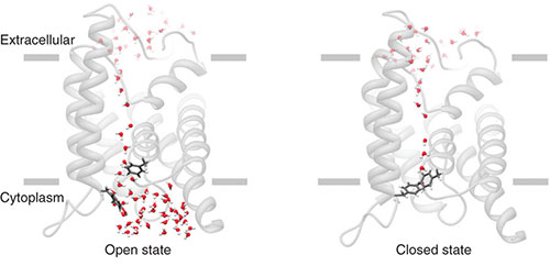
Close-up view of water pores in the eye's lens

NIH study of aquaporins could hold clues to genetic errors in eye structure, damage to eyes from diabetes and smoking, as well as the advance of cataracts in aging eyes.

Researchers have achieved dynamic, atomic-scale views of a protein needed to maintain the transparency of the lens in the human eye. The work, funded in part by the National Institutes of Health, could lead to new insights and drugs for treating cataract and a variety of other health conditions.


Aquaporin proteins form water channels between cells and are found in many tissues, but aquaporin zero (AQP0) is found only in the mammalian lens, which focuses light onto the retina, at the back of the eye. The lens is primarily made up of unique cells called lens fibers that contain little else besides water and proteins called crystallins. Tight packing of these fibers and of the crystallin proteins within them helps create a uniform medium that allows light to pass through the lens, almost as if it were glass.


Abnormal development or age-related changes in the lens can lead to cataract—a clouding of the lens that causes vision loss. Besides age, other risk factors for cataract include smoking, diabetes, and genetic factors. Mutations in the AQP0 gene can cause congenital cataract and may increase the risk of age-related cataract.

"The AQP0 channel is believed to play a vital role in maintaining the transparency of the lens and in regulating water volume in the lens fibers, so understanding the molecular details of how water flows through the channel could lead to a better understanding of cataract," said Dr. Houmam Araj, who oversees programs on lens, cataract and oculomotor systems at NIH’s National Eye Institute (NEI), which helped fund the research.


Closing of AQP0 channels is regulated by a calcium-sensitive protein called calmodulin, but the precise mechanism has been unclear. Some models have suggested that calmodulin simply acts as a plug to fill the open channel. The new study, published in Nature Structural and Molecular Biology, reveals a more nuanced process in which calmodulin essentially grasps the open channel and forces it to close.


The research was a collaboration between investigators at the University of California, Irvine and the Janelia Farm Research Campus in Ashburn, Va., a part of the Howard Hughes Medical Institute (HHMI). Drs. James Hall and Douglas Tobias led the effort at UC Irvine. Dr. Tamir Gonen led the effort at Janelia Farm.

In prior studies, Dr. Gonen had examined the atomic structure of the AQP0 protein by X-ray crystallography, which involves crystallizing a protein and bombarding it with X-rays. But X-ray crystallography does not work well for large groups of proteins or for proteins in motion. So in the new study, the researchers first used electron microscopy to view AQP0 and calmodulin bound together. Then they combined their microscopy and crystallography data to generate computerized models of how the two proteins interact and to identify the most critical amino acids (the building blocks for proteins) within AQP0. To test their models, they neutralized those amino acids one-by-one in the actual AQP0 channel.


The AQP0 channel is made up of four identical barrel-shaped units, bundled together side by side. The researchers found that in the presence of calcium, calmodulin binds to one unit and then another, as if grabbing a pair of reins. This makes the channel twist slightly, which causes just a few amino acids within each unit to slide into the channel’s core and block the flow of water.


"Calmodulin essentially throws a molecular switch that moves in and out of the water pore, like the gate valve of a plumbing fixture," Dr. Hall said.

This new view of AQP0 could help lead to new approaches for treating cataract, Dr. Hall said. Cataracts are the most common cause of blindness worldwide. In the U.S., they affect about one in six people over age 40 and half over age 80. Congenital cataracts (present from birth) affect about 1 in 5000, American children.

Cataracts can be successfully treated with surgery, in which the cloudy lens is removed and replaced with an artificial plastic lens. But the new findings "may be a step toward learning how to prevent or delay cataracts," said Dr. Hall.

The study, by Reichow SL, Clemens DM, Freites JA, Németh-Cahalan KL, Heyden M, Tobias DJ, Hall JE, and Gonen T., is titled: "Allosteric mechanism of water-channel gating by Ca2+–calmodulin." and available in Nature Structural and Molecular Biology, July 2013. DOI: 10.1038/nsmb.2630.

The new findings also provide inroads to understanding how calmodulin interacts with a variety of protein channels, and thus could open doors to new drugs for other common health conditions. In addition to aquaporins, our bodies rely on a vast menagerie of channels, many of which are regulated by calmodulin. For example, calmodulin helps control the gating of ion channels, which allow the passage of ions (charged particles) in and out of our cells and are essential for nerve cell firing, muscle contraction, and the rhythmic beating of the heart. This study provides the first structural model of calmodulin bound to any complete protein channel.

Abstract
Calmodulin (CaM) is a universal regulatory protein that communicates the presence of calcium to its molecular targets and correspondingly modulates their function. This key signaling protein is important for controlling the activity of hundreds of membrane channels and transporters. However, understanding of the structural mechanisms driving CaM regulation of full-length membrane proteins has remained elusive. In this study, we determined the pseudoatomic structure of full-length mammalian aquaporin-0 (AQP0, Bos taurus) in complex with CaM, using EM to elucidate how this signaling protein modulates water-channel function. Molecular dynamics and functional mutation studies reveal how CaM binding inhibits AQP0 water permeability by allosterically closing the cytoplasmic gate of AQP0. Our mechanistic model provides new insight, only possible in the context of the fully assembled channel, into how CaM regulates multimeric channels by facilitating cooperativity between adjacent subunits.

Drs. Daniel Clemens and Steve Reichow were co-first authors on the study. NIH support for the study came from NEI (grants EY005661, EY018768), the National Institute of General Medical Sciences (NIGMS grant GM079233), a joint program on "Making Sense of Voltage Sensors" co-funded by NIGMS and the National Institute of Neurological Disorders and Stroke (grant GM086685), and the National Library of Medicine (grant LM007443). Additional support came from HHMI, the National Science Foundation, and the German Academy of Sciences.

To set up an interview with Dr. Hall, please contact Tom Vasich at UC Irvine (tmvasich@uci.edu, 949-824-6455).

The National Eye Institute, part of the National Institutes of Health, leads the federal government's research on the visual system and eye diseases. NEI supports basic and clinical science programs that result in the development of sight-saving treatments. For more information, visit http://www.nei.nih.gov.

About the National Institutes of Health (NIH): NIH, the nation's medical research agency, includes 27 Institutes and Centers and is a component of the U.S. Department of Health and Human Services. NIH is the primary federal agency conducting and supporting basic, clinical, and translational medical research, and is investigating the causes, treatments, and cures for both common and rare diseases. For more information about NIH and its programs, visit http://www.nih.gov.

NIH...Turning Discovery Into Health®

Original press release:http://www.nei.nih.gov/news/pressreleases/080213.asp