|
|
|
Home | Pregnancy Timeline | News Alerts |News Archive Oct 9, 2014
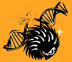
  Illustration by the journal Developmental Cell
When the protein Boc is inactivated in early medulloblastoma, tumor numbers dropped 66 percent. |
|
 |
|
|
|
Bad signal to Sonic Hedgehog creates brain tumors
A protein named Boc is needed to signal the gene Sonic Hedgehog to begin cell divisions resulting in very specific patterns that become very specific structures. But an error in Boc over stimulates Sonic Hedgehog to over promote growth of cerebellum brain cells leading to medulloblastoma in children.
Medulloblastoma ranks among the leading causes of cancer-related death in children. Current treatment includes surgery with radiation and chemotherapy. Although the majority of children survive treatment, radiation therapy damages normal brain cells in infants and toddlers causing long-term problems.
Scientists at the Institute of Research Clinics of Montreal (IRCM) in Canada have discovered a mechanism that promotes the progression of medulloblastoma, the most common brain tumor found in children. A team led by Frédéric Charron, PhD, found that the protein known as Boc can send a faulty signal to Sonic Hedgehog inducing it to spur cell proliferation leading to brain cancer.
This important breakthrough will be published in the October issue of the prestigious scientific journal Developmental Cell. The editors also selected the article to be featured on the journal’s cover.
Sonic Hedgehog (Shh) belongs to a family of proteins that direct cell location and patterning in early embryo development. Faulty regulation of Shh plays a significant role in tumor genesis, the process which changes normal cells into cancer cells.
“Our team studied a protein called Boc, a receptor located on the cell surface that binds to Sonic Hedgehog. We had previously shown that Boc is important in the development of the cerebellum, the part of the brain where medulloblastomas arise, so we decided to investigate its role further,” explains Lukas Tamayo-Orrego, PhD student in Dr. Charron’s laboratory and co-first author of the study.
Adds Dr. Charron, Director of the Molecular Biology of Neural Development research unit at the IRCM:“With this study, we identified that the presence of Boc is required for Sonic Hedgehog. Sonic Hedgehog then drives the proliferation of granule precursor cells (GCPs) (granule cells are found within the granular layer of the cerebellum, the dentate gyrus of the hippocampus, the superficial layer of the dorsal cochlear nucleus, the olfactory bulb, and the cerebral cortex). An error in activating Sonic Hedgehog can induce an overproliferation of GCPs [granule precursor cells], leading to medulloblastoma. In fact, Boc itself can cause DNA mutation, promoting the progression of precancerous lesions to become advanced medulloblastoma.”
"Our study shows that when Boc is inactivated, the number of tumors is reduced by 66 per cent. The inactivation of Boc therefore reduces the development of early medulloblastoma into advanced tumors,” says Frederic Mille, PhD, co-first author of the article and former postdoctoral fellow in Dr. Charron’s research unit.
“Our results indicate that Boc could potentially be targeted to develop a new therapeutic approach to stop the growth and progression of medulloblastoma and thus reduce the adverse side effects of current treatment for medulloblastomas.”
Frédéric Charron PhD, Director, Molecular Biology of Neural Development research, Institute of Research Clinics of Montreal, Canada, co-first author of the research presented.
Article Summary
During cerebellar development, Sonic hedgehog (Shh) signaling drives the proliferation of granule cell precursors (GCPs). Aberrant activation of Shh signaling causes overproliferation of GCPs, leading to medulloblastoma. Although the Shh-binding protein Boc associates with the Shh receptor Ptch1 to mediate Shh signaling, whether Boc plays a role in medulloblastoma is unknown. Here, we show that BOC is upregulated in medulloblastomas and induces GCP proliferation. Conversely, Boc inactivation reduces proliferation and progression of early medulloblastomas to advanced tumors. Mechanistically, we find that Boc, through elevated Shh signaling, promotes high levels of DNA damage, an effect mediated by CyclinD1. High DNA damage in the presence of Boc increases the incidence of Ptch1 loss of heterozygosity, an important event in the progression from early to advanced medulloblastoma. Together, our results indicate that DNA damage promoted by Boc leads to the demise of its own coreceptor, Ptch1, and consequently medulloblastoma progression.
About the research project
This research project was supported by grants from the Canadian Institutes of Health Research, the Canadian Cancer Society and the Cancer Research Society. Other authors from the IRCM include Martin Lévesque (co-first author), Julie Cardin, Nicolas Bouchard, and Luisa Izzi. The project was also conducted in collaboration with the laboratories of Stefan Pfister in Heidelberg, Germany, and Michael Taylor in Toronto.
About the IRCM
The IRCM (www.ircm.qc.ca) is a renowned biomedical research institute located in the heart of Montréal’s university district. Founded in 1967, it is currently comprised of 35 research units and four specialized research clinics (cholesterol, cystic fibrosis, diabetes and obesity, hypertension). The IRCM is affiliated with the Université de Montréal, and the IRCM Clinic is associated to the Centre hospitalier de l’Université de Montréal (CHUM). It also maintains a long-standing association with McGill University. The IRCM is funded by the Quebec ministry of Economy, Innovation and Export Trade (Ministère de l’Économie, de l’Innovation et des Exportations).
Return to top of page |
