|
|
|
Home | Pregnancy Timeline | News Alerts |News Archive Nov 27, 2014
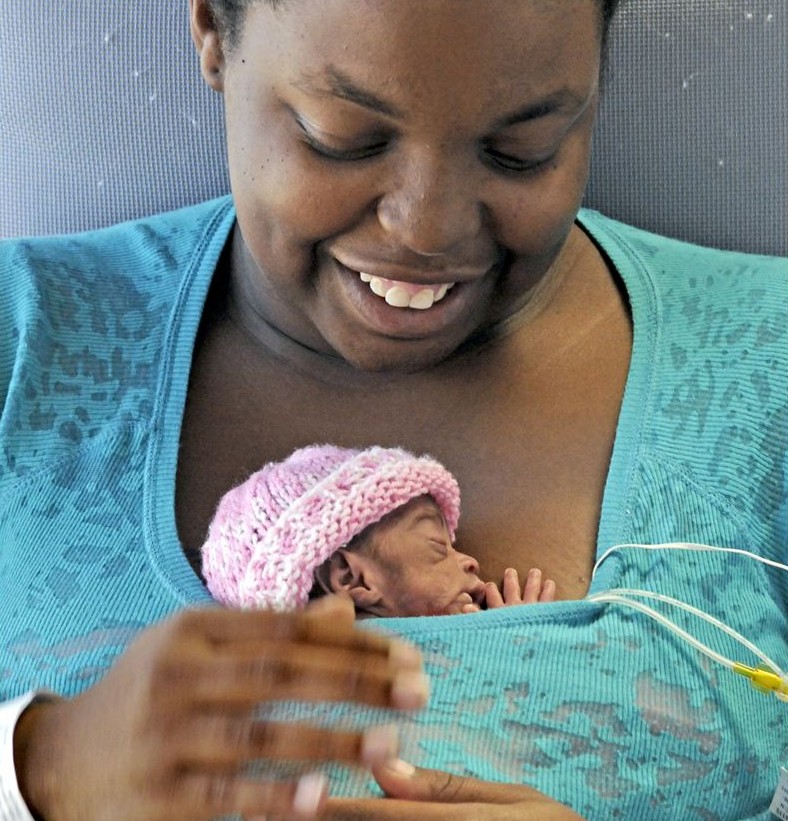
A mother's touch offers insight into maternal caregiving as it affects a developing brain. |
|
|
|
|
|
Mom's presence changes genes in infant brain
A mother's tender loving care, her "TLC", can not only help soothe infants in pain, but may actually impact early brain development by altering gene activity in the amygdala.
According to research led by New York University (NYU), Langone Medical Center, a loving mother's touch can create positive effects on a developing brain.
By carefully analyzing infant rat brains, NYU researchers found that several hundred genes became active when pups experienced a painful stimulus. However, with their mothers present, fewer than 100 genes were similarly expressed in pups' brains under the same stimulus.
According to Regina Sullivan PhD, senior investigator and neurobiologist who led the research, this is the first time that short-term affects of maternal caregiving have been reflected in a distressed pup's brain. The study was designed to reflect the long-term consequences of maternal nuturing in mammals — beginning with birth.
"Our study shows that a mother's ability to comfort an infant in pain does not just elicit a behavioral response, but can also modify critically developing neural circuitry in the early brain."
Regina Sullivan PhD, professor, NYU School of Medicine, Nathan S. Kline Institute for Psychiatric Research.
Dr. Sullivan's work is published in Current Biology.
Sullivan's previous research has revealed that a rat mother's physical presence affects the electrical signals being sent through an infant pup's brain. With this research, her analysis of gene changes in amygdalas of infant rats — a brain region responsible for processing fear, pain and pleasure — adds to the complexity of understanding how pain in newborns, whether rats or humans, affects the developing brain.
"Nobody wants to see an infant suffer, in rats or any species," says Sullivan. "But, if opiates are too dangerous to use in human infants because of their addictive properties, the challenge remains to find alternatives — including maternal comfort, coddling, or cues such as a mother's scent, that might relieve infant pain and protect the developing brain."
"The more we learn about the infant brain, the better we can treat pain from extensive surgeries or any physical or mental abuse experienced in infancy."
Regina Sullivan PhD
Highlights
•The mother’s presence reduces infant rat cortical desynchronization
•Maternal behaviors (e.g., milk ejection and grooming) increase desynchronization
•Maternal effects on infant cortical activity decline with age
•Norepinephrine receptor blockade reduces impact of dam on infant cortical activity
Summary
Patterns of neural activity are critical for sculpting the immature brain, and disrupting this activity is believed to underlie neurodevelopmental disorders [ 1–3 ]. Neural circuits undergo extensive activity-dependent postnatal structural and functional changes [ 4–6 ]. The different forms of neural plasticity [ 7–9 ] underlying these changes have been linked to specific patterns of spatiotemporal activity. Since maternal behavior is the mammalian infant’s major source of sensory-driven environmental stimulation and the quality of this care can dramatically affect neurobehavioral development [ 10 ], we explored, for the first time, whether infant cortical activity is influenced directly by interactions with the mother within the natural nest environment. We recorded spontaneous neocortical local field potentials in freely behaving infant rats during natural interactions with their mother on postnatal days ∼12–19. We showed that maternal absence from the nest increased cortical desynchrony. Further isolating the pup by removing littermates induced further desynchronization. The mother’s return to the nest reduced this desynchrony, and nipple attachment induced a further reduction but increased slow-wave activity. However, maternal simulation of pups (e.g., grooming and milk ejection) consistently produced rapid, transient cortical desynchrony. The magnitude of these maternal effects decreased with age. Finally, systemic blockade of noradrenergic beta receptors led to reduced maternal regulation of infant cortical activity. Our results demonstrate that during early development, mother-infant interactions can immediately affect infant brain activity, in part via a noradrenergic mechanism, suggesting a powerful influence of the maternal behavior and presence on circuit development.
Besides Sullivan, Gordon A. Barr, at the University of Pennsylvania, served as lead investigator for this study.
Funding support for the study was provided by the US National Institute of Child Health and Human Development and the National Institute of Mental Health, both parts of the National Institutes of Health. Corresponding federal grant numbers are R01 DC009910 and R01 MH0901451.
Return to top of page |


