|

CLICK ON weeks 0 - 40 and follow along every 2 weeks of fetal development
|
||||||||||||||||||||||||||||
|
|
|||||||||||||||||||||||||||||
|
Home | Pregnancy Timeline | News Alerts | News Archive May 16, 2013
|
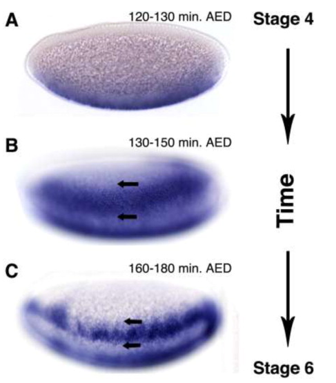
The developmental genetics of space and time Understanding the mechanism by which a signal from one part of an embryo can influence the location and other variables of surrounding cells—is important to gene regulation, evolution, and human health. by Albert Erives Researchers have conducted a study that reveals important and useful insights into how and why developmental genes often take inputs from two independent “morphogen concentration gradients.”Albert Erives, associate professor in the University of Iowa Department of Biology, led the work. His graduate student, Justin Crocker, is currently a postdoctoral researcher at the Howard Hughes Medical Institute (HHMI) Janelia Farm Research Campus. The study appears in the Genomes & Developmental Control section of the online June 1 issue of the journal Developmental Biology. Understanding the concept of morphogen gradients—the mechanism by which a signal from one part of a developing embryo can influence the location and other variables of surrounding cells—is important to developmental biology, gene regulation, evolution, and human health. Morphogen gradients subdivide a field of cells into territories characterized by distinct cell fates. These divisions allow cells to “know” their position within a developing embryo and to differentiate appropriate to that location. However, in order to function, such systems require a genetic mechanism to regulate a spectrum of responses to different genes. This mechanism is called a transcriptional enhancer. These are DNA sequences that display a cryptic code of transcription factor (TF) binding sites. During development and sometimes under environmental influences, these enhancers assemble scaffolds for TF protein complexes that orchestrate differential gene expression. However, enhancers targeted by morphogen signals may temporally initate inappropriate gene expression as the morphogen builds up and decays over a specific window of the embryo's developmental timeline. Using the Drosophila (fruit fly) gene system, which includes many species—each with a fully sequenced genome—the Erives Lab identified a case of spatial and temporal (time) conflict in the regulation of the ventral neurons defective (vnd) gene. The vnd gene must be precisely regulated in order for the fly’s nervous system to be properly developed. VND is turned on by a key embryonic factor (dorsal/NFkB) that creates the pattern of dorsal/ventral axis in an embryo. In particular, the vnd gene plays a critical role in specifying distinct dorsal/ventral neural columns in the ectoderm by repressing additional regulators. In this regulatory hierarchy, vnd requires early expression, but ventral neurogenic ectoderm demands a relatively high-threshold response to the morphogen. However, the study also shows that the vnd gene’s Neurogenic Ectoderm Enhancer (NEE) can take input from a complementary gradient of the Dpp morphogen via the Schnurri/Mad/Medea silencer element (SSE). In this regard, the NEE at vnd is unlike NEEs at other genetic loci, which are not involved in the neural specification circuit and have no resident SSE. The study also shows that an SSE could be added to a single-input NEE and cause spatial restriction of its activity. These results show how requirements for conflicting temporal and spatial responses to one morphogen can be solved by adding input from complementary morphogens. The Erives Lab at the UI’s Department of Biology studies the structure, function, and evolution of enhancers within the context of gene regulatory circuits underlying the evolution and development of animals by using molecular, genetic, and evolutionary genomic approaches. Within these areas, the Erives Lab has published several landmark papers notable for demonstrating how whole genome sequences can be used to accelerate biological research on outstanding questions in biology. Original article: http://now.uiowa.edu/2013/05/developmental-genetics-space-and-time |
||||||||||||||||||||||||||||