|
|
|
Home | Pregnancy Timeline | News Alerts |News Archive Nov 13, 2014
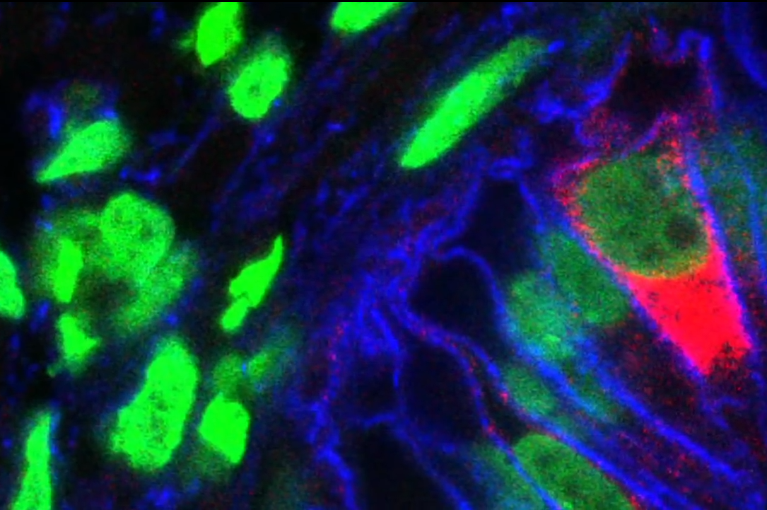
Gastric Organoids made from pluripotent stem cells mimic human stomach epithelium.
Image credit: Cincinnati Children's Hospital
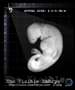
Inset: Carnegie Stage 13 Digestive epithelium layer begins to differentiate into the future
locations for the liver, lung, stomach and pancreas.
Image inset credit: The Visible Embryo |
|
|
|
|
|
Human stomach created from pluripotent stem cells
For the first time, scientists using pluripotent stem cells, have generated functioning, three-dimensional, human stomach tissue in a laboratory. This work has created a new tool for studying how tissues develop and how infection and disease create change.
Scientists at Cincinnati Children's Hospital Medical Center reported on Oct. 29 in Nature magazine that they were able to use human pluripotent stem cells – cells that can become any cell type in the body – to grow miniature versions of the human stomach. Collaborating with researchers at the University of Cincinnati College of Medicine, they generated mini-stomachs — or gastric organoids — in order to study infection by the bacteria H. pylori, a major cause of peptic ulcers and stomach cancer.
This first-time creation of 3D human gastric organoids (hGOs) presents opportunities to model early stage stomach cancer and study obesity related diabetes. It also is the first time research has produced a 3D human embryonic foregut – a promising starting point for generating other organ tissues made from the foregut – such as lungs and pancreas.
"Until this study, no one had generated gastric cells from human pluripotent stem cells (hPSCs). We've now discovered how to stimulate formation of three-dimensional gastric tissue with a complex architecture and cellular composition."
Jim Wells PhD, principal investigator, division of Developmental Biology and Endocrinology, Cincinnati Children's Hospital.
There are many subtle differences between our species and the mouse, which is often used to model humans in research. And the architecture of the human stomach makes mouse models less than ideal for the study of human stomach diseases.
Researchers can now use human gastric organoids as a tool to help identify biochemical processes in the gut which allow gastric-bypass patients to become diabetes-free soon after surgery even before losing significant weight.
Diabetes and metabolic syndrome are exploding epidemics. Until now, a major challenge to investigating these conditions has been a lack of reliable animal models that accurately simulate human biology.
The key to growing human gastric organoids was to identify the steps involved in stomach formation during embryonic development. By manipulating normal processes in a petri dish, the scientists were able to coax pluripotent human stem cells into becoming stomach cells. Over the course of a month, these steps resulted in the formation of 3D gastric organoids of about 3mm (1/10th of an inch) in diameter. Wells and his colleagues were then able to identify what drives normal stomach formation in humans, their intention being to understand what goes wrong when the stomach does not properly form.
In one experiment they were impressed by how rapidly the H. pylori bacteria infected stomach epithelial tissues. Within 24 hours, H. pylori had triggered biochemical changes in the stomach cells — and the human gastric organoids faithfully mimicked these same early stages of gastric disease. The H. pylori bacteria activated a cancer gene called c-Met as the bacterial infection rapidly spread throughout the epithelial tissues.
"This milestone work would not have been possible if it hadn't been for studies in basic research to understand embryonic organ development."
Jim Wells PhD
Abstract
Gastric diseases, including peptic ulcer disease and gastric cancer, affect 10% of the world’s population and are largely due to chronic Helicobacter pylori infection1, 2, 3. Species differences in embryonic development and architecture of the adult stomach make animal models suboptimal for studying human stomach organogenesis and pathogenesis4, and there is no experimental model of normal human gastric mucosa. Here we report the de novo generation of three-dimensional human gastric tissue in vitro through the directed differentiation of human pluripotent stem cells. We show that temporal manipulation of the FGF, WNT, BMP, retinoic acid and EGF signalling pathways and three-dimensional growth are sufficient to generate human gastric organoids (hGOs). Developing hGOs progressed through molecular and morphogenetic stages that were nearly identical to the developing antrum of the mouse stomach. Organoids formed primitive gastric gland- and pit-like domains, proliferative zones containing LGR5-expressing cells, surface and antral mucous cells, and a diversity of gastric endocrine cells. We used hGO cultures to identify novel signalling mechanisms that regulate early endoderm patterning and gastric endocrine cell differentiation upstream of the transcription factor NEUROG3. Using hGOs to model pathogenesis of human disease, we found that H. pylori infection resulted in rapid association of the virulence factor CagA with the c-Met receptor, activation of signalling and induction of epithelial proliferation. Together, these studies describe a new and robust in vitro system for elucidating the mechanisms underlying human stomach development and disease.
Funding support for the research came in part from the National Institutes of Health (R01DK080823, R01DK092456, K01DK091415), the NIGMS Medical Scientist Training Program (T32 GM063483), the American Gastroenterological Association: Robert and Sally Funderburg Research Award in Gastric Cancer, the Cincinnati Digestive Disease Center Award (P30 DK0789392), Clinical Translational Science Award (U54 RR025216), and the Michigan Gastrointestinal Peptide Research Center (MGPRC; NIDDK 5P30DK034933).
About Cincinnati Children's:
Cincinnati Children's Hospital Medical Center ranks third in the nation among all Honor Roll hospitals in U.S.News and World Report's 2014 Best Children's Hospitals. It is also ranked in the top 10 for all 10 pediatric specialties. Cincinnati Children's, a non-profit organization, is one of the top three recipients of pediatric research grants from the National Institutes of Health, and a research and teaching affiliate of the University of Cincinnati College of Medicine. The medical center is internationally recognized for improving child health and transforming delivery of care through fully integrated, globally recognized research, education and innovation. Additional information can be found at http://www.cincinnatichildrens.org. Connect on the Cincinnati Children's blog, via Facebook and on Twitter.
Return to top of page
|