|
|
Welcome to The Visible Embryo, a comprehensive educational resource on human development from conception to birth.
The Visible Embryo provides visual references for changes in fetal development throughout pregnancy and can be navigated via fetal development or maternal changes.
The National Institutes of Child Health and Human Development awarded Phase I and Phase II Small Business Innovative Research Grants to develop The Visible Embryo. Initally designed to evaluate the internet as a teaching tool for first year medical students, The Visible Embryo is linked to over 600 educational institutions and is viewed by more than one million visitors each month.
Today, The Visible Embryo is linked to over 600 educational institutions and is viewed by more than 1 million visitors each month. The field of early embryology has grown to include the identification of the stem cell as not only critical to organogenesis in the embryo, but equally critical to organ function and repair in the adult human. The identification and understanding of genetic malfunction, inflammatory responses, and the progression in chronic disease, begins with a grounding in primary cellular and systemic functions manifested in the study of the early embryo.

The World Health Organization (WHO) has created a new Web site to help researchers, doctors and patients obtain reliable information on high-quality clinical trials. Now you can go to one website and search all registers to identify clinical trial research underway around the world!

|
|
| Disclaimer: The Visible Embryo web site is provided for your general information only. The information contained on this site should not be treated as a substitute for medical, legal or other professional advice. Neither is The Visible Embryo responsible or liable for the contents of any websites of third parties which are listed on this site. |
|
|

Content protected under a Creative Commons License. Commons License. |
|
| No dirivative works may be made or used for commercial purposes. |
|
|
| |
|
|
 
CLICK ON weeks 0 - 40 and follow along every 2 weeks of fetal development
|
|
|
|
Fetal Timeline Maternal Timeline News News Archive Sep 8, 2015
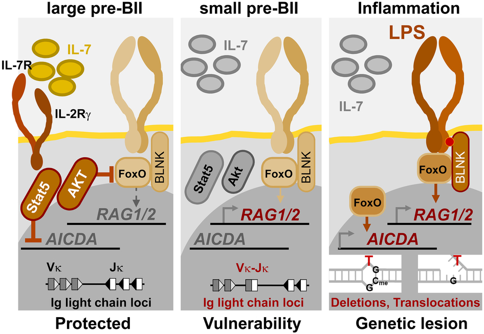
Schematic: Loss of IL-7R signaling at small pre-BII makes a pre-B cell vulnerable to
acquisition of genetic changes by activation of AID, RAG1 and RAG2.
Image Credit: UCSF .
|
|
| |
|
|
|
Childhood Vaccination Reduces Leukemia Risk
Research has discovered how a commonly given vaccine protects against acute lymphoblastic leukemia (ALL), the most common type of childhood cancer.
Adapted from an article by Juliana Bunim | UCSF.edu
The Haemophilus influenzae Type b (Hib) vaccine not only prevents ear infections and meningitis caused by the bacteria, but also protects against ALL, or approximately 25 percent of cancer diagnoses among children younger than 15 years old, according to the National Cancer Society. The Hib vaccine is part of the standard vaccination schedule recommended by the Centers for Disease Control and is routinely given to children in four doses before they are 15 months old.
Though the cancer protection offered by the Hib vaccine has been well established in epidemiological studies, it is not well-known among the public.
Perhaps because the mechanism underlying this effect has been poorly understood and therefore poorly explained by physicians.
Reported in Nature Immunology, an international team led by University of California San Francisco (UCSF) researchers has shown that recurrent Hib infections can put certain immune-system genes into overdrive, converting “pre-leukemia” blood cells — which are present in a surprisingly large number of newborns — into full-blown cancer.
“The incidence of leukemia has been dramatically reduced since the advent of regular HIB vaccinations during infancy. Hib and other childhood infections can cause recurrent and vehement immune responses, of which some were found to lead to leukemia. But most infants that received vaccines are protected and acquire long-term immunity through very mild immune reactions.”
Markus Müschen, MD, PhD, Professor of Laboratory Medicine, UCSF and senior author of the study.
Many newborns carry oncogenes — or genes that could potentially cause cancer — in their blood cells, but only one in 10,000 will eventually develop ALL. Oncogenes may be the result of the instability of newly developing blood cells. In the new study, researchers tested the idea that chronic inflammation caused by recurrent infections might cause “collateral damage” — or additional genetic lesions — in blood cells already carrying an oncogene, promoting their transformation to overt disease.
Led by co-first authors Srividya Swaminathan, PhD, a former UCSF postdoctoral fellow now at Stanford University School of Medicine, and Lars Klemm, assistant research specialist at UCSF, the team conducted experiments with mice that homed in on two enzymes known as AID and RAG as the drivers of this process.
AID and RAG introduce DNA mutations that allow immune cells to adapt to infection. These two enzymes are needed for a normal and efficient immune response. But in the presence of chronic infection, AID and RAG become hyperactivated, randomly cutting and mutating genes, including important gatekeepers against cancer.
By studying genetically engineered pre-leukemia cells lacking either AID or RAG, or both, the team found AID and RAG working together is critical to introduce the additional lesions that result in life-threatening disease.
Abstract
Childhood acute lymphoblastic leukemia (ALL) can often be traced to a pre-leukemic clone carrying a prenatal genetic lesion. Postnatally acquired mutations then drive clonal evolution toward overt leukemia. The enzymes RAG1-RAG2 and AID, which diversify immunoglobulin-encoding genes, are strictly segregated in developing cells during B lymphopoiesis and peripheral mature B cells, respectively. Here we identified small pre-BII cells as a natural subset with increased genetic vulnerability owing to concurrent activation of these enzymes. Consistent with epidemiological findings on childhood ALL etiology, susceptibility to genetic lesions during B lymphopoiesis at the transition from the large pre-BII cell stage to the small pre-BII cell stage was exacerbated by abnormal cytokine signaling and repetitive inflammatory stimuli. We demonstrated that AID and RAG1-RAG2 drove leukemic clonal evolution with repeated exposure to inflammatory stimuli, paralleling chronic infections in childhood.
Return to top of page
|