|
|
Developmental Biology - Cell Structure
Cell Cytoskeleton Is a Permanent Construction Site
A cell's cytoskeleton consists of protein filaments that are continually lengthening and shortening in dynamic tank like movement...
Cell changes in shape and movement occur throughout cell division, differentiation into new cellular identities and processes such as wound healing.
If something goes wrong in the cytoskeletal construction site - such as protein filaments that have undergone remodeling at the wrong place or time - disease can ensue. Such errors can initiate metastatic cancer cells to migrate.
Researchers at the Max Delbrück Center for Molecular Medicine in the Helmholtz Association (MDC) and the European Bioinformatics Institute (EMBL-EBI) in Hinxton, United Kingdom, have investigated how a family of 145 proteins causes these remodeling events to occur at the right place and time. Up to now, scientists had only examined these regulatory proteins in individual studies, and only a few proteins had been characterized.
"In order to understand complex processes, including cell shape changes, we need to know how regulatory proteins work together."
Oliver Rocks PhD, Max Delbrück Center for Molecular Medicine, Berlin, Germany; Lunenfeld-Tanenbaum Research Institute, Sinai Health System, Toronto, Canada.
The study is published in the journal Nature Cell Biology. Lead authors are Dr. Paul M. Müller and Dr. Juliane Rademacher with Dr. Evangelia Petsalaki and her group at EMBL-EBI, along with an international research team. All regulatory proteins have now systematically been characterized.
The team is now able to show how different signaling zones in a cell coordinate the cytoskeleton in space and time as well as how zones are created and maintained.
A New Perspective, Thanks to a Comprehensive Database
At the cytoskeletal construction site, Rho GTPase proteins set the tone. When these molecular switches are activated, they send commands to the molecular machinery. There are 145 regulatory proteins that control these molecular switches with RhoGEF proteins turning them on, and RhoGAP proteins turning them off.
Oliver Rocks and his team have systematically investigated all these regulators for the first time and created a kind of library. Researchers all over the world can access this library to see which molecular switches individual proteins control, where in the cell this occurs and which binding partners it has.
The comprehensive information contained in the protein library allows proteins to be analyzed at the systems level for the first time - i.e., from a bird's eye perspective. This has revealed new collective properties of the regulators that were previously imperceptible. In this way, researchers discovered a new mechanism that explains how cell migration is controlled.
Focal Adhesions Control Balance of Opposing Processes
Scientists were aware that two opposing processes control the cytoskeleton - cell protrusion and cell contraction - need to occur in separate areas in the cell to allow for cellular movement. In one construction site at the front of the cell, molecular switches command the cytoskeleton protrusion toward the migration direction. At the cell interior, molecular switches trigger contraction of the cytoskeleton. The MDC team investigated the central question of how Rho GTPases coordinates the two spatially separated processes.
They found cell spatial organization of two opposing processes:
(1) focal adhesions located close to the front of the cell mature and eventually dissolve.
(2) focal adhesions located slightly back and directly below the cell membrane, anchor the cell to its environment
(3) when travelling, focal adhesions move from front to middle of the cell.
"The cell exploits the fact that focal adhesions change location. The real surprise is that we found a specific subgroup of regulators located almost exclusively on newly formed focal adhesions at the cell's edge and a separate subgroup on mature structures towards the middle of the cell."
Oliver Rocks PhD, Max Delbrück Center for Molecular Medicine, Berlin, Germany; Lunenfeld-Tanenbaum Research Institute, Sinai Health System, Toronto, Ontario, Canada.
These subgroups control opposing cytoskeletal processes, creating spatially separated signal zones. Researchers were able to show both processes are linked by mechanical forces in the cell, and help maintain balance between the number of newly formed focal adhesions and the number of mature focal adhesions (which dissipate).
Rocks next plans to investigate how precisely Rho GTPase regulators of focal adhesions communicate with cytoskeletal machinery. The question being how or whether the principle of spatial separation between these proteins plays a role in other cell structures; and how and if, a defect in regulatory function may lead to disease.
This systemic view of cell shape regulation "...was absolutely essential to revitalize this research field and open up new conceptual approaches for further study," explains Rocks.
The database and protein library are now available to all scientists worldwide.
Abstract
County-level ASD prevalence was estimated using an age-resolved snapshot from the California Department of Developmental Services (DDS) for birth years 19932013. ASD prevalence increased among all children across birth years 19932000 but plateaued or declined thereafter among whites from wealthy counties. In contrast, ASD rates increased continuously across 19932013 among whites from lower income counties and Hispanics from all counties. Both white ASD prevalence and rate of change in prevalence were inversely correlated to county income from birth year 20002013 but not 19932000. These disparate trends within the dataset suggest that wealthy white parents, starting around 2000, may have begun opting out of DDS in favor of private care and/or making changes that effectively lowered their childrens risk of ASD.
Authors
Cynthia Nevison and William Parker.
Acknowledgements
The authors thank the California Department of Developmental Services (CDDS) for providing the data used in this study. We are grateful to Sallie Bernard and Irva Hertz-Picciotto for their helpful comments. We also thank two anonymous reviewers, whose comments much improved the manuscript. Finally, we thank Susanne Meza-Keuthen for her support of this study.
Funding
None of the authors received specific funding for this work.
Return to top of page.
| |
|
Mar 27 2020 Fetal Timeline Maternal Timeline News
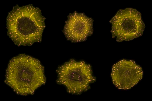 Microscopic images of cells in which filaments of the actin cytoskeleton are labelled in yellow and focal adhesions labelled in pink. CREDIT Markus Müller, Rocks Lab, MDC.
|