|
|
Developmental Biology - Gene Therapy
Measuring Cancer Cell Mechanics in Living Animals
Researchers now have the ability to measure and quantify nanomechanical factors in living animal cells...
A first of its kind nanoparticle based in vivo (living creature) imaging technique may one day be used to help diagnose and even treat cancer. The technique was developed by collaborating researchers from Michigan State University (MSU), Johns Hopkins University (JHU) and Stanford University.
Research results are published in the journal Materials Today. Bryan Smith PhD, associate professor of biomedical engineering at MSU, worked with colleagues to develop the tiny particles, which placed inside living cells, can reveal important information about cell structure - including how tumor cells physically change as they form a tumor.
"We engineered the ability to measure and quantify the nanomechanical properties of individual living cells within the body of a living animal for the first time."
Bryan Smith PhD, Associate Professor, Biomedical Engineering, Michigan State University, USA.
In a study earlier this year, Smith and his team designed nanoparticles that helped "eat" away atherosclerosis, the plaque buildup in arteries that can lead to heart attack. The particles selectively entered immune system cells known as macrophages, delivering a drug instructing cells to devour the harmful plaques.
Now, Smith and his colleagues have created a technique using different nanoparticles that can be embedded into various cell types, including cancerous breast cells, in live animals. Analyzing how these particles move within the cell can reveal a lot about its internal physical properties. They plan to look at the formation and dissemination of cancer metastases, which cause about 90% of cancer deaths.
"There previously existed no method to examine mechanical properties in living subjects - for example, in mammals - with high spatial resolution. Such techniques promise to open entirely new avenues of inquiry for both disease diagnosis and treatment."
Bryan Smith PhD
The mechanical properties of biological tissues have been known to play a major role in many disease states, including heart disease, inflammation and cancer, as well as normal physiology such as cell migration and organism development. In the current study, Smith and his team used nanoparticles to first compare the mechanical properties between cells in culture - both standard 2D and 3D - and in live animals.
Tracking movement of nanoparticles revealed that their environment greatly affects their mechanical properties - which could mean that certain cell models may not be valid representations of live animals.
"This tells cancer scientists interested in cancer mechanics that 2D conditions may poorly replicate, and that certain 3D conditions be substantially closer in mimicking conditions within the live mouse,"
Bryan Smith PhD
The next part of the experiment looked at what actually happens to the internal structure of cancer cells as they begin to form tumors. Previous methods couldn't answer the question because they were too invasive to test in living subjects. Again, after observing the movement of the nanoparticles within the cells, the team measured how "compliant" or soft, these cells were. Importantly, they found how normal cells' pliability remained steady over time, but as cancer cells formed into a tumor over the period of a week, they stiffened.
"We found that as a tumor begins to form in a living mouse, individual tumor cells mechanically stiffen. This is a fundamental finding which is ultimately likely to have implications for cancer spread (metastasis) and tumor lethality. The discovery was made possible by integrating state-of-the-art imaging and particle tracking technologies from our and our collaborators' labs."
Bryan Smith PhD
The research has a number of promising applications in medicine. One of these is simply evaluating which cell culture methods are enough like living organisms to provide meaningful information. Another is measuring the cell mechanical properties of common biological functions, including organ development, in living organisms.
Perhaps the most exciting application may be in disease diagnosis and treatment according to Smith. Nanoparticles might be used to monitor the health of cells and the types of changes they undergo in disease processes - and may even alter that course.
"I hope one day we'll be able to treat the physics of metastasis. But, we must first understand the mechanics and how changing them impacts cell behavior."
Bryan Smith PhD, Associate Professor, Biomedical Engineering at MSU
Abstract
Cumulative evidence shows that microenvironmental conditions play a significant role in the regulation of cell functions, and how cells respond to these conditions are of central importance to regenerative medicine and cancer cell response to therapeutics. Here, we develop a new method to examine cell mechanical properties by analyzing the motion of nanoparticles in living in mice, combining particle tracking with intravital microscopy. This method directly examines the mechanical response of breast carcinoma cells and normal breast epithelial cells under intravital microenvironments. Our results show both carcinoma and normal cells display significantly reduced compliance (less deformability) in vivo compared to the same cells cultured in 2D, in both sparse and confluent conditions. While the compliance of the normal cells remains steady over time, the compliance of carcinoma cells decreases further as they form tumor-like architectures. Integrating the cancer cells into spheroids embedded in 3D collagen matrices in part redirected the mechanical response to a state closer to the in vivo setting. Overall, our study demonstrates that the microenvironment is a crucial regulator of cell mechanics and the intravital particle tracking method can provide novel insights into the role of cell mechanics in vivo.
Authors
Pei-Hsun Wu, Sanjiv Sam Gambhir, Christopher M. Hale, Wei-Chiang Chen, Denis Wirtz, Bryan Ronain Smith.
Return to top of page.
| |
|
May 4 2020 Fetal Timeline Maternal Timeline News
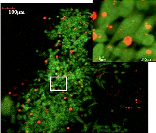 Early tumor (GREEN) cells with (RED) nanoparticles within them. Tumor cells were measured by their mechanical properties using microrheology. This is the first image from a video taken within a living mouse. The inset shows individual tumor cells and nanoparticles at higher resolution. CREDIT Bryan Smith
|