|
|
|
Home | Pregnancy Timeline | News Alerts |News Archive Mar 17, 2015
UMMS scientists have developed a multicolored CRISPR/Cas9 labeling system that precisely
measures the distance between two points on one chromosome, as well as the location
of genes on a chromosome, and where in the nucleus the chromosome resides.
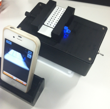
Image: Arizona State University created a DNA fluorimeter APP for smartphones. |
|
|
|
|
|
How to build a gene location APP
There is a new "app" for finding and mapping specific gene locations on a gene or DNA sequence of a chromosome. Scientists from the University of Massachusetts Medical School (UMMS) are using one of the hottest tools in biomedical research to locate genes on chromosomes — CRISPR/Cas9.
CRISPR stands for Clustered Regularly Interspersed Palindromic Repeats — a portion of DNA that remains the same when it is reversed. Together with the enzyme Cas9, CRISPR/Cas9 becomes a gene-editing machine revolutionizing genome engineering — in use only since 2013. Scientists use RNA guides to select gene locations on DNA, and then using CRISPR/Cas9, cut out that particular DNA sequence. After Cas9 cuts the target sequence, the cell is forced to repair the damage using a slightly altered version provided by a scientist.
Because it is trivial to make an RNA guide to direct Cas9 to where to cut a gene, CRISPR tremendously simplifies the process of deleting, adding, or modifying genes. As of 2014 it had successfully been tested in 20 species, including humans. In many of those species, edits were made in cells that give rise to sperm or eggs, allowing future generations in that species to inherit the alteration.
The CRISPR/CAS9 labeling system could become key to manipulating gene expression as it allows researchers to measure the precise distances between two known points on different chromosomes within living cells.
The work is published in PNAS, and was first presented at the American Society for Cell Biology/International Federation for Cell Biology annual meeting in Philadelphia in December 2014.
The nucleus of every cell in our bodies (with the exception of eggs, sperm and red blood cells) must pack in 23 pairs of chromosomes (46 individual ones). Tightly wound bundles of extremely long strands of DNA are wrapped around protein knobs known as histones. For a gene to be transcribed and produce a protein, it must become slightly unwound from these histones to be accessible and be "read" by cell machinery.
Scientists have long suspected that the position a chromosome holds within the nucleus also affects it's gene accessibility. So knowing the location of the chromosome within the nucleus as well as the formation of chromosomes is critical to understanding how genes actually work. The human nucleus is very crowded, says study authors Thoru Pederson, PhD, professor of biochemistry and molecular pharmacology, and research specialist Hanhui Ma, PhD, at the University of Massachusetts Medical School.
By deploying pairs of fluorescent tags from a three-color labeling system that their lab developed, Pederson and colleagues show that it is possible to plot not only where a chromosome is within the cell nucleus, but where it is relative to other chromosomes.
Their CRISPR app can also measure the distance between two points on one chromosome, providing an idea of it's level of compaction — key to gene expression of a protein.
Precisely locating chromosomes in the nucleus of living cells has been a holy grail in cell biology since Joseph Gall and Mary Lou Pardue first detected specific loci in 1968. Their discovery opened the era of genetic testing, but early testing required "fixed" or dead cells.
In the intervening years, research has created new methods to probe live cells including transcription activator-like effectors (TALEs), which Ma and Pederson recently introduced. TALEs light up genetic loci in living cells. But, both now feel the rapidly emerging CRISPR system promises a more accurate map of a living nucleus while being easier to employ.
Using their multicolored labeling system, Ma and Pederson were able to determine locations of several chromosomes. Among them, gene-rich chromosome 19 tends to be located in the middle of the nucleus, whereas gene-poor chromosome 18 is on the periphery.
Also staying close to the center of the nucleus are chromosome 17 and five of the so-called acrocentric chromosomes, which have distinctive centromeres close to the ends of one short arm. [If the short arm of a chromosome is so short that it is hard to observe, but still present, then that chromosome is acrocentric ("acro-" is the Greek word for "peak"). The human genome includes five acrocentric chromosomes: 13, 14, 15, 21, 22. Wikipedia]
One of the five acrocentrics is chromosome 21. An extra copy of chromosome 21 is called trisomy 21 and is the diagnostic marker for Down syndrome.
Meanwhile, chromosomes 3 and 7 are at, or close to, the periphery of the nucleus.
Ma and Pederson were also able to distinguish different locations for each diploid copy (two complete sets of chromosomes, one from each parent) of genes involved in organizing the nucleolus. They have plans to further tweak their two-color technique to find translocations — the abnormal switches in chromosome segments found in human tumor cells.
Ultimately, the authors believe the GPS app will provide scientists a new toolkit to "study the 4D nucleome and regulation of gene expression in a broad landscape of cell types and stages of development, differentiation and disease."
Significance
The detection of specific genes in fixed cells was first accomplished in 1969 by Gall and Pardue. The development of analogous methods applicable to living cells is now at hand. At the forefront of this advance (2013–2014), we and other investigators have used transcription activator-like effectors (TALEs) conjugated with fluorescent proteins to tag genomic loci in live cells. More recently, the CRISPR/Cas9 system has provided a more flexible approach to targeting specific loci. In this paper, we describe the labeling of human genomic loci in live cells with three orthogonal CRISPR/Cas9 components, allowing multicolor detection of genomic loci with high spatial resolution, which provides an avenue for barcoding elements of the human genome in the living state.
Abstract
The intranuclear location of genomic loci and the dynamics of these loci are important parameters for understanding the spatial and temporal regulation of gene expression. Recently it has proven possible to visualize endogenous genomic loci in live cells by the use of transcription activator-like effectors (TALEs), as well as modified versions of the bacterial immunity clustered regularly interspersed short palindromic repeat (CRISPR)/CRISPR-associated protein 9 (Cas9) system. Here we report the design of multicolor versions of CRISPR using catalytically inactive Cas9 endonuclease (dCas9) from three bacterial orthologs. Each pair of dCas9-fluorescent proteins and cognate single-guide RNAs (sgRNAs) efficiently labeled several target loci in live human cells. Using pairs of differently colored dCas9-sgRNAs, it was possible to determine the intranuclear distance between loci on different chromosomes. In addition, the fluorescence spatial resolution between two loci on the same chromosome could be determined and related to the linear distance between them on the chromosome’s physical map, thereby permitting assessment of the DNA compaction of such regions in a live cell.
The published study was funded in part by the U.S. National Science Foundation.
About the University of Massachusetts Medical School
The University of Massachusetts Medical School (UMMS), one of five campuses of the University system, comprises the School of Medicine, the Graduate School of Biomedical Sciences, the Graduate School of Nursing, a thriving research enterprise and an innovative public service initiative, Commonwealth Medicine. Its mission is to advance the health of the people of the commonwealth through pioneering education, research, public service and health care delivery with its clinical partner, UMass Memorial Health Care. In doing so, it has built a reputation as a world-class research institution and as a leader in primary care education. The Medical School attracts more than $240 million annually in research funding, placing it among the top 50 medical schools in the nation. In 2006, UMMS's Craig C. Mello, PhD, Howard Hughes Medical Institute Investigator and the Blais University Chair in Molecular Medicine, was awarded the Nobel Prize in Physiology or Medicine, along with colleague Andrew Z. Fire, PhD, of Stanford University, for their discoveries related to RNA interference (RNAi). The 2013 opening of the Albert Sherman Center ushered in a new era of biomedical research and education on campus. Designed to maximize collaboration across fields, the Sherman Center is home to scientists pursuing novel research in emerging scientific fields with the goal of translating new discoveries into innovative therapies for human diseases.
Return to top of page
|


