Welcome to The Visible Embryo
  o
Home-- -History-- -Bibliography- -Pregnancy Timeline- --Prescription Drugs in Pregnancy- -- Pregnancy Calculator- --Female Reproductive System- -Contact

Welcome to The Visible Embryo, a comprehensive educational resource on human development from conception to birth.

The Visible Embryo provides visual references for changes in fetal development throughout pregnancy and can be navigated via fetal development or maternal changes.

The National Institutes of Child Health and Human Development awarded Phase I and Phase II Small Business Innovative Research Grants to develop The Visible Embryo. Initally designed to evaluate the internet as a teaching tool for first year medical students, The Visible Embryo is linked to over 600 educational institutions and is viewed by more than one million visitors each month.

Today, The Visible Embryo is linked to over 600 educational institutions and is viewed by more than 1 million visitors each month. The field of early embryology has grown to include the identification of the stem cell as not only critical to organogenesis in the embryo, but equally critical to organ function and repair in the adult human. The identification and understanding of genetic malfunction, inflammatory responses, and the progression in chronic disease, begins with a grounding in primary cellular and systemic functions manifested in the study of the early embryo.

WHO International Clinical Trials Registry Platform


The World Health Organization (WHO) has created a new Web site to help researchers, doctors and
patients obtain reliable information on high-quality clinical trials. Now you can go to one website and search all registers to identify clinical trial research underway around the world!


Home

History

Bibliography

Pregnancy Timeline

Prescription Drug Effects on Pregnancy

Pregnancy Calculator

Female Reproductive System

Contact The Visible Embryo

News Alerts Archive

Disclaimer: The Visible Embryo web site is provided for your general information only. The information contained on this site should not be treated as a substitute for medical, legal or other professional advice. Neither is The Visible Embryo responsible or liable for the contents of any websites of third parties which are listed on this site.
Content protected under a Creative Commons License.

No dirivative works may be made or used for commercial purposes.


Pregnancy Timeline by SemestersLungs begin to produce surfactantImmune system beginningHead may position into pelvisFull TermPeriod of rapid brain growthWhite fat begins to be madeHead may position into pelvisWhite fat begins to be madeImmune system beginningBrain convolutions beginBrain convolutions beginFetal liver is producing blood cellsSensory brain waves begin to activateSensory brain waves begin to activateInner Ear Bones HardenBone marrow starts making blood cellsBone marrow starts making blood cellsBrown fat surrounds lymphatic systemFetal sexual organs visibleFinger and toe prints appearFinger and toe prints appearHeartbeat can be detectedHeartbeat can be detectedBasic Brain Structure in PlaceThe Appearance of SomitesFirst Detectable Brain WavesA Four Chambered HeartBeginning Cerebral HemispheresFemale Reproductive SystemEnd of Embryonic PeriodEnd of Embryonic PeriodFirst Thin Layer of Skin AppearsThird TrimesterSecond TrimesterFirst TrimesterFertilizationDevelopmental Timeline
CLICK ON weeks 0 - 40 and follow along every 2 weeks of fetal development
Google Search artcles published since 2007


Home | Pregnancy Timeline | News Alerts |News Archive Mar 24, 2015

Testing about 20 human lymphoma and leukemia B cell lines in 2013, researchers
found that most can be transdifferentiated at least partially into macrophage-like cells,
provided that C/EBPα is expressed at sufficiently high levels.
Image Credit: the journal Cell


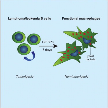
Leukemia cells changed into harmless immune cells

After a chance observation in the lab, researchers at Stanford University School of Medicine found found they could change dangerous leukemia cells into mature and harmless immune cells called macrophages.

The findings are described in the paper published online March 16 in the Proceedings of the National Academy of Sciences.

B-cell acute lymphoblastic leukemia with a mutation called the Philadelphia chromosome is a particularly aggressive cancer with poor outcomes, according to Ravi Majeti MD PhD an assistant professor of medicine and senior author on the paper.

Majeti and his colleagues made the observation after collecting leukemia cells from a patient and attempting to keep the cells alive culture. "We were throwing everything at them to help them survive," said Majeti, who is also a member of the Stanford Cancer Institute and the Stanford Institute for Stem Cell Biology and Regenerative Medicine.

Postdoctoral scholar Scott McClellan MD, PhD, lead author on the paper, mentioned that some of the cancer cells in culture were changing shape and size into what looked like macrophages. Majeti agreed, but why the cells changed was a mystery. Then he recalled a research paper which showed early B-cell mouse progenitor cells could be forced to become macrophages when exposed to certain transcription factors — proteins that bind to certain DNA sequences.


"B-cell leukemia cells are in many ways progenitor cells that are forced to stay in an immature state."

Ravi Majeti MD PhD, Assistant Professor, Medicine, Stanford Cancer Institute and the Stanford Institute for Stem Cell Biology and Regenerative Medicine, and senior author.


So McClellan and student Christopher Dove, MD/PhD student and the paper's second lead author, conducted more experiments confirming that methods developed years ago could be used to transform human cancer cells into macrophages, which engulf and digest cancer cells and pathogens.

Majeti and his colleagues hoped to find that after cancer cells become macrophages they not only become neutralized, but may actually assist in fighting cancer. Macrophage cells present recognizable bits of abnormal cells to other immune cells so those cells can launch an attack. "Because the macrophage cells initially came from those cancer cells, they will already carry within them chemical signals to identify them as cancer cells, making an immune attack against the cancer more likely," Majeti theorized.

Researchers will next look for a drug that will prompt the same reaction and serve as the basis for an ALL leukemia therapy. There is precedent for such a treatment as Retinoic acid is commonly used to treat acute promyelocytic leukemia after it was found that Retinoic acid turns cancer cells into mature cells called granulocytes. This is the only well-established therapy to mature or differentiate cancer cells. But researchers around the world are hopeful of finding many more.

Majeti: "There's big-time interest in differentiation therapies for cancer."

Significance
Precursor B cell acute lymphoblastic leukemia (B-ALL) is an aggressive cancer of white blood cells with a poor prognosis. The cancerous cells in this disease are immature B cells, which are unable to fully differentiate into normal B cells. We show here that cancerous cells from B-ALL patients can be reprogrammed, causing them to change into cells that resemble normal macrophages and can perform macrophage-associated functions such as the consumption of bacteria. Importantly, unlike typical B-ALL cells, these reprogrammed cells are no longer able to cause disease in immunodeficient mice. Finally, we show that this reprogramming process may occur to some degree in patients with B-ALL. This indicates that reprogramming B-ALL cells into macrophages might represent a previously unidentified therapeutic strategy.

Abstract
BCR–ABL1+ precursor B-cell acute lymphoblastic leukemia (BCR–ABL1+ B-ALL) is an aggressive hematopoietic neoplasm characterized by a block in differentiation due in part to the somatic loss of transcription factors required for B-cell development. We hypothesized that overcoming this differentiation block by forcing cells to reprogram to the myeloid lineage would reduce the leukemogenicity of these cells. We found that primary human BCR–ABL1+ B-ALL cells could be induced to reprogram into macrophage-like cells by exposure to myeloid differentiation-promoting cytokines in vitro or by transient expression of the myeloid transcription factor C/EBPα or PU.1. The resultant cells were clonally related to the primary leukemic blasts but resembled normal macrophages in appearance, immunophenotype, gene expression, and function. Most importantly, these macrophage-like cells were unable to establish disease in xenograft hosts, indicating that lineage reprogramming eliminates the leukemogenicity of BCR–ABL1+ B-ALL cells, and suggesting a previously unidentified therapeutic strategy for this disease. Finally, we determined that myeloid reprogramming may occur to some degree in human patients by identifying primary CD14+ monocytes/macrophages in BCR–ABL1+ B-ALL patient samples that possess the BCR–ABL1+ translocation and clonally recombined VDJ regions.

Majeti is a New York Stem Cell Foundation Robertson Investigator.

Other Stanford co-authors of the paper are computational biologist Andrew Gentles, PhD, and technician Christine Ryan, who is now a medical student at Stanford.

This research was supported by the National Institutes of Health (grant U54CA149145), the New York Stem Cell Foundation, the Burroughs Wellcome Fund, the U.S. Department of Defense and the Walter V. and Idun Berry Postdoctoral Fellowship Program.

The Stanford University School of Medicine consistently ranks among the nation's top medical schools, integrating research, medical education, patient care and community service. For more news about the school, please visit http://med.stanford.edu/school.html. The medical school is part of Stanford Medicine, which includes Stanford Health Care and Lucile Packard Children's Hospital Stanford. For information about all three, please visit http://med.stanford.edu.

Return to top of page