Autism's early 'neighborhood'
Scientists have found that in children with autism, sensorimotor regions of the brain become overconnected at the expense of later-developing higher-order brain functions.
In early childhood, neurons inside a child's developing brain form connections between various regions of brain "real estate." As described in a paper published last week in the journal Biological Psychiatry, cognitive neuroscientists at San Diego State University (SDSU) found sensorimotor regions of the brain appear to be overdeveloped in children and adolescents with autism spectrum disorder.
Overdevelopment appears to infringe on brain "real estate" that in typically developing children is more densely occupied by connections to serve higher cognitive functioning.
The study represents the first ever systematic look at connections between the entire cerebral cortex and the cerebellum using fMRI brain imaging. Its findings provide another piece in the puzzle that could one day lead to development of a brain-based test identifying autism.
Several decades ago, scientists reported findings that certain regions of the cerebellum — a brain region involved in motor control, but also in cognitive, social, and emotional functions — were often smaller in people with autism than in typically developing people.
That sparked a brief flurry of research activity exploring the cerebellum's potential role in the disorder. Unfortunately, the direction never truly panned out for researchers hoping for a big breakthrough in understanding, said the study's corresponding author, SDSU psychologist Ralph-Axel Müller.
"Eventually, interest in the cerebellum waned due to a lack of consistency in the findings."
Ralph-Axel Müller, psychologist, San Diego State University
Hoping that advances in brain imaging technology would reveal new insights, Müller, worked with the study's first author Amanda Khan and looked back at the cerebellum. Khan is a former master's student at SDSU and now a doctoral candidate at Suffolk University in Boston.
They directed 56 children and adolescents, half with autism and half without, to fixate on a focal point while thinking about nothing in particular. Using fMRI brain imaging technology, they scanned the children's brains during this activity. Capturing this spontaneous activity is crucial to honing in on what are essentially baseline neural patterns.
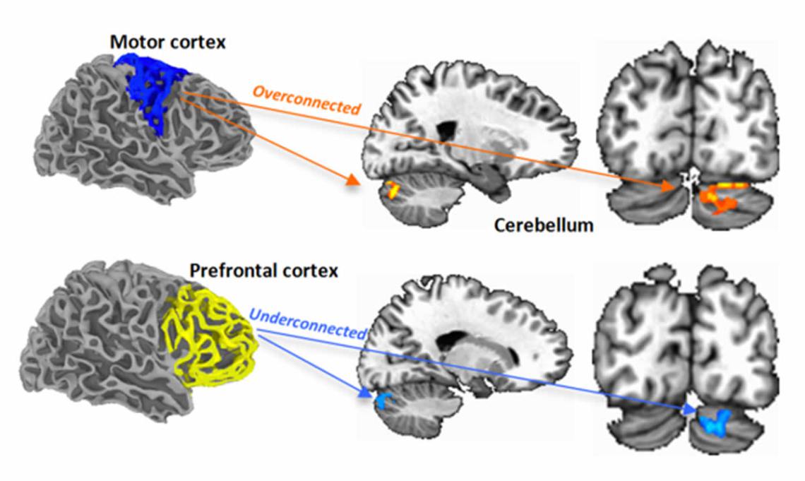
The imaging results revealed that participants with autism had far stronger neuronal connectivity between sensorimotor regions of the cerebellum and cerebral cortex than did their counterparts without autism. Conversely, participants with autism had less connectivity between regions involved in higher-order cognitive functions such as decision-making, attention and language.
Müller explained that sensorimotor connections between the cerebral cortex and cerebellum mature during the first few years of life, when the brains of children with autism grow larger in volume than typically developing children. Connections that serve higher cognitive functions develop later, after this period of overgrowth.
"Our findings suggest that the early developing sensorimotor connections are highly represented in the cerebellum at the expense of higher cognitive functions in children with autism.
"By the time the higher cognitive functions begin to come online, many of the connections are already specialized. If a particular part of the brain is already functionally active in one domain, there may be no reason for the brain to switch it over to another domain later in life."
Ralph-Axel Müller
Returning to the real estate metaphor, it was as if most of the available land has already been scooped up by sensorimotor connections before the higher-order cognitive function connections have a chance to move into the neighborhood.
These findings could help scientists and clinicians better understand exactly how abnormalities during brain development lead to various types of autism spectrum disorder.
Müller hopes his work will not only contribute to a brain-based diagnosis of autism, but also be a step towards identifying its various subtypes and underlying genetic factors.
Müller: "We still don't understand what in the brain makes a kid autistic. You can't look at a scan and say, 'There it is.' However, we're doing the groundwork of finding brain variables that might be biomarkers for autism and its subtypes."
Abstract
Background
The cerebellum plays important roles in both sensorimotor and supramodal cognitive functions. Cellular, volumetric, and functional abnormalities of the cerebellum have been found in autism spectrum disorders (ASD), but no comprehensive investigation of cerebro-cerebellar connectivity in ASD is available.
Methods
We used resting-state functional connectivity MRI in 56 children and adolescents (28 ASD, 28 typically developing [TD]) aged 8-17 years. Partial and total correlation analyses were performed for unilateral regions of interest (ROIs), distinguished in two broad domains as sensorimotor (premotor/primary motor, somatosensory, superior temporal, occipital) and supramodal (prefrontal, posterior parietal, and inferior and middle temporal).
Results
There were three main findings: (i) Total correlation analyses showed predominant cerebro-cerebellar functional overconnectivity in the ASD group; (ii) partial correlation analyses that emphasized domain-specificity (sensorimotor vs. supramodal) indicated a pattern of robustly increased connectivity in the ASD group (compared to the TD group) for sensorimotor ROIs, but predominantly reduced connectivity for supramodal ROIs; (iii) this atypical pattern of connectivity was supported by significantly increased non-canonical connections (between sensorimotor cerebral and supramodal cerebellar ROIs, and vice versa) in the ASD group.
Conclusions
Our findings indicate that sensorimotor intrinsic functional connectivity is atypically increased in ASD, at the expense of connectivity supporting cerebellar participation in supramodal cognition.
SDSU graduate students Aarti Nair and Christopher Keown, also contributed to the study, as did University of California, San Diego, graduate student Michael Datko and Alliant International University psychologist Alan Lincoln.
About San Diego State University
San Diego State University is a major public research institution offering bachelor's degrees in 89 areas, master's degrees in 78 areas and doctorates in 21 areas. The university provides transformative experiences, both inside and outside of the classroom, for its 34,000 students. Students participate in research, international experiences, sustainability and entrepreneurship initiatives, and a broad range of student life and leadership opportunities. The university's rich campus life features opportunities for students to participate in, and engage with, the creative and performing arts, a Division I athletics program and the vibrant cultural life of the San Diego region. For more information, visit http://www.sdsu.edu.
Return to top of page
|