|
|
Welcome to The Visible Embryo, a comprehensive educational resource on human development from conception to birth.
The Visible Embryo provides visual references for changes in fetal development throughout pregnancy and can be navigated via fetal development or maternal changes.
The National Institutes of Child Health and Human Development awarded Phase I and Phase II Small Business Innovative Research Grants to develop The Visible Embryo. Initally designed to evaluate the internet as a teaching tool for first year medical students, The Visible Embryo is linked to over 600 educational institutions and is viewed by more than one million visitors each month.
Today, The Visible Embryo is linked to over 600 educational institutions and is viewed by more than 1 million visitors each month. The field of early embryology has grown to include the identification of the stem cell as not only critical to organogenesis in the embryo, but equally critical to organ function and repair in the adult human. The identification and understanding of genetic malfunction, inflammatory responses, and the progression in chronic disease, begins with a grounding in primary cellular and systemic functions manifested in the study of the early embryo.

The World Health Organization (WHO) has created a new Web site to help researchers, doctors and patients obtain reliable information on high-quality clinical trials. Now you can go to one website and search all registers to identify clinical trial research underway around the world!

|
|
| Disclaimer: The Visible Embryo web site is provided for your general information only. The information contained on this site should not be treated as a substitute for medical, legal or other professional advice. Neither is The Visible Embryo responsible or liable for the contents of any websites of third parties which are listed on this site. |
|
|
 |
|
Content protected under a Creative Commons License. Commons License.
No dirivative works may be made or used for commercial purposes. |
|
|
| |
|
|

CLICK ON weeks 0 - 40 and follow along every 2 weeks of fetal development
|
|
|
|
|
Home | Pregnancy Timeline | News Alerts |News Archive Apr 30, 2015
|
|
|
Link between physical forces and limb deformities
Engineers and a pediatric surgeon have joined forces to discover that physical forces like pressure and tension affect the development of limbs in embryos — research that could someday be used to help prevent birth defects.
The team included University of Toronto (U of T) mechanical engineer Yu Sun, Department of Mechanical and Industrial Engineering (MIE), University of Toronto, Canada, and bioengineer Rodrigo Fernandez-Gonzalez, assistant professor at the Institute of Biomaterials & Biomedical Engineering (IBBME), and Dr. Sevan Hopyan of the SickKids Hospital.
Their study — published in Nature Cell Biology — used live imaging and computer models to gain valuable insight into how mechanical forces change cell shape and cell movement into arms and legs. A mouse embryo begins shaped like a ball, then expands and creates the complex shapes of limbs. But first, as an early embryo, its cells divide into three layers:
ectoderm: forms the nervous system, skin and sensory organs
mesoderm: produces skeleton, muscle and most major organs
endoderm: becomes respiratory tract and elimination systems
In the study, the team looked at cell behavior in the ectoderm layer that also promotes limb development. Using unique tools, including micro-chiseling lasers, atomic force microscopes and layer-by-layer computer modeling, they explored the early stages of limb bud growth in unprecedented detail.
Researchers discovered that as cells divide and develop, they not only communicate with each other, but are impacted by the pressures from movement within each of the other cell layers. All cummulatively impacting limb bud formation.
Prior to these experiments, scientists and engineers didn't have the tools and techniques to understand how change of shape on the molecular scale impacts cells as they form into tissues. But thanks to this work, researchers now know that the ectoderm and mesoderm speak to each other not only biochemically by shuttling molecules back and forth between cell walls, but also mechanically through cell mass redistribution.
"The idea that two tissues are mechanically interacting and that such interaction affects cellular behavior is really exciting to see," says Fernandez-Gonzalez.
To measure mechanical forces, the authors used techniques borrowed from the world of manufacturing and engineering, including the use of a laser to cut between cell walls.
"We know some of the genes that are important in the structure of the embryo for development to proceed, but we didn't know how those pathways were linked with movement in the cells."
Sevan Hopyan MD, PhD, FRCSC, Assistant Professor
Department of Surgery and Department of of Molecular Genetics, The Hospital for Sick Children (SickKids) with the University of Toronto, Canada.
The team foresees future 'measurement of movement' simulations in cell remodeling using their technique and tools. "This research could someday also be used in potential medical applications to prevent limb deformations," says Dr. Hopyan.
Abstract
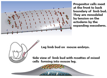
The physical forces that drive morphogenesis are not well characterized in vivo, especially among vertebrates. In the early limb bud, dorsal and ventral ectoderm converge to form the apical ectodermal ridge (AER), although the underlying mechanisms are unclear. By live imaging mouse embryos, we show that prospective AER progenitors intercalate at the dorsoventral boundary and that ectoderm remodels by concomitant cell division and neighbour exchange. Mesodermal expansion and ectodermal tension together generate a dorsoventrally biased stress pattern that orients ectodermal remodelling. Polarized distribution of cortical actin reflects this stress pattern in a β-catenin- and Fgfr2-dependent manner. Intercalation of AER progenitors generates a tensile gradient that reorients resolution of multicellular rosettes on adjacent surfaces, a process facilitated by β-catenin-dependent attachment of cortex to membrane. Therefore, feedback between tissue stress pattern and cell intercalations remodels mammalian ectoderm.
Return to top of page
|