Welcome to The Visible Embryo
  o
Home-- -History-- -Bibliography- -Pregnancy Timeline- --Prescription Drugs in Pregnancy- -- Pregnancy Calculator- --Female Reproductive System- -Contact
 

Welcome to The Visible Embryo, a comprehensive educational resource on human development from conception to birth.

The Visible Embryo provides visual references for changes in fetal development throughout pregnancy and can be navigated via fetal development or maternal changes.

The National Institutes of Child Health and Human Development awarded Phase I and Phase II Small Business Innovative Research Grants to develop The Visible Embryo. Initally designed to evaluate the internet as a teaching tool for first year medical students, The Visible Embryo is linked to over 600 educational institutions and is viewed by more than one million visitors each month.

Today, The Visible Embryo is linked to over 600 educational institutions and is viewed by more than 1 million visitors each month. The field of early embryology has grown to include the identification of the stem cell as not only critical to organogenesis in the embryo, but equally critical to organ function and repair in the adult human. The identification and understanding of genetic malfunction, inflammatory responses, and the progression in chronic disease, begins with a grounding in primary cellular and systemic functions manifested in the study of the early embryo.

WHO International Clinical Trials Registry Platform


The World Health Organization (WHO) has created a new Web site to help researchers, doctors and patients obtain reliable information on high-quality clinical trials. Now you can go to one website and search all registers to identify clinical trial research underway around the world!


Home

History

Bibliography

Pregnancy Timeline

Prescription Drug Effects on Pregnancy

Pregnancy Calculator

Female Reproductive System

Contact The Visible Embryo

News Alerts Archive

Disclaimer: The Visible Embryo web site is provided for your general information only. The information contained on this site should not be treated as a substitute for medical, legal or other professional advice. Neither is The Visible Embryo responsible or liable for the contents of any websites of third parties which are listed on this site.
Content protected under a Creative Commons License.

No dirivative works may be made or used for commercial purposes.


Pregnancy Timeline by SemestersDevelopmental TimelineFertilizationFirst TrimesterSecond TrimesterThird TrimesterFirst Thin Layer of Skin AppearsEnd of Embryonic PeriodEnd of Embryonic PeriodFemale Reproductive SystemBeginning Cerebral HemispheresA Four Chambered HeartFirst Detectable Brain WavesThe Appearance of SomitesBasic Brain Structure in PlaceHeartbeat can be detectedHeartbeat can be detectedFinger and toe prints appearFinger and toe prints appearFetal sexual organs visibleBrown fat surrounds lymphatic systemBone marrow starts making blood cellsBone marrow starts making blood cellsInner Ear Bones HardenSensory brain waves begin to activateSensory brain waves begin to activateFetal liver is producing blood cellsBrain convolutions beginBrain convolutions beginImmune system beginningWhite fat begins to be madeHead may position into pelvisWhite fat begins to be madePeriod of rapid brain growthFull TermHead may position into pelvisImmune system beginningLungs begin to produce surfactant
CLICK ON weeks 0 - 40 and follow along every 2 weeks of fetal development


Google Search artcles published since 2007
   

Home | Pregnancy Timeline | News Alerts |News Archive Jun 3, 2015


(ABOVE) Drop-seq technique

(BELOW) in-Drops technique


Why and how to barcode thousands of cells

When it comes to the tissues in our bodies, which cells do what, is almost always misleading. Scientists know there isn't just one cell type in an organ or any tissue.


Scientists run tests to determine what molecules are in their samples. This is useful information, but it doesn't tell where those molecules originally came from. It only provides an average of the type of cells within that sample.


"If you take a hunk of tissue and grind it up and analyze the RNA, you have no idea if it represents what every cell in that population is doing or what no cell in that population is doing."

Marc Kirschner, the John Franklin Enders University Professor of Systems Biology and chair of the Department of Systems Biology at Harvard Medical School.


The trouble is, it's expensive, time-consuming and tricky to characterize tissues one cell, or cell type, at a time.

Kirschner and Steven McCarroll, assistant professor of genetics at Harvard Medical School (HMS), now report In separate papers — McCarrol in the journal Cell1 and Kirschner also in the journal Cell2 — that each of their labs has developed a system to identify individual cells. Each lab announced a high-throughput technique to quickly and inexpensively assign unique genetic barcodes to every cell in a sample.

As a result, scientists can analyze complex tissues by profiling each individual cell without having to average all cells.

"Different cells in a tissue use the same genome in amazingly diverse ways: to engineer specialized cell shapes, accomplish diverse feats of physiology, and mount distinct functional responses to the same stimulus. These techniques will finally let science understand how biological systems operate at that single-cell level," said McCarroll, who is also director of genetics for the Stanley Center for Psychiatric Research at the Broad Institute of Harvard and MIT.


To make their tools, both teams collaborated with David Weitz, Mallinckrodt Professor of Physics and Applied Physics at Harvard's School of Engineering and Applied Sciences, and a pioneer in the field of microfluidics.


The teams expect that each of their techniques will allow biologists to classify cell types and map cell diversity in complex tissues such as those found in the brain. Harvard's Office of Technology Development has been working closely with the researchers to develop patent applications with an eye toward commercializing each method.


Gene expression is the activity of a gene or genes in a particular cell; it underlies all biological function from thinking to development of the egg. Scientists knew for 50 years that gene expression varied from cell to cell. But they haven't been able to expediently measure a single-cell in a tissue sample as tissues include many cell types, in different stages of cell division in any sample.


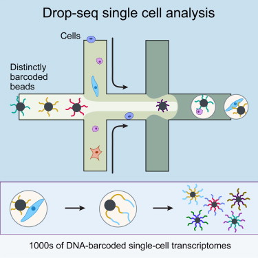
But now, Evan Macosko PhD in the McCarroll lab, a Stanley Neuroscience Fellow and HMS instructor in psychiatry at Massachusetts General Hospital, has developed a technique he calls "Drop-seq".

Alternatively, Klein, assistant professor of systems biology at HMS on the Kirschner team, has devised a method for indexing droplets by sequencing, called "inDrops".

Each method uses tiny beads to simultaneously deliver vast numbers of unique DNA barcodes into hundreds of thousands of nanometer-sized water droplets. Thanks to David Weitz's expertise in applied physics and with his assistance, both methods use microfluid devices to encapsulate cells with beads inside of each water droplet. The droplets get created in a tiny assembly line, streaming along a channel the width of a human hair.

Bead barcodes attach to each gene within a cell. This allows scientists to sequence each gene in a tissue sample while still being able to trace that gene back to its original cell.

However, Macosko and Klein make their beads differently. Their droplets get broken up at different steps in their unique processes and each uses diverging aspects of chemistry. But, their results are the same.

After running a single batch of cells through "Drop-seq" or "inDrops", scientists can see which genes are expressed in the entire sample — and can sort by each individual cell," says Klein. They can then use computer software to uncover patterns in the overall mix of cells, including which cells have similar gene profiles. This allows scientists to classify which cell types appeared in a sample — and even discover new ones.


Current methods allows research to generate 96 single-cell expression profiles a day for several thousand dollars.

Drop-seq, by comparison, enables 10,000 profiles a day for 6.5 cents each.


Macosko adds: "It finally makes gene expression profiling on a cell-by-cell level, tractable and accessible. I think it's something biologists in a lot of fields will want to use."

Rather than competing with each other, the teams believe that having two options available with "Drop-seq" and "inDrops" will benefit the scientific community. "Each method has unique elements that makes it better for different applications. Biologists will be able to choose which one is most appropriate for them," said Macosko.

McCarroll, Macosko and colleagues are excited to explore the brain with Drop-seq. This will include the search for new cell types, constructing a global architecture of all brain cell types, and lead to a better understanding of brain development and function as related to disease. Among questions they want to pursue: (1) What are all of the cell types making up the brain? (2) How do these cell types vary in function and response to stimuli? (3) What cell populations are missing or malfunctioning in schizophrenia, autism and other brain disorders?

Immediately, Sanes is completing a catalog of cell types in the mouse retina. Drop-seq has already revealed several new ones.


Classifying cell types may not sound exciting but it is the foundation for mapping neural circuits and probing the mystery of how the brain gives rise to thoughts, emotions and behaviors.


Meanwhile, Kirschner, Klein and colleagues are keenly interested in stem cell development.

"Does a population of cells that we initially think is uniform actually have some substructure?" asks Allon Klein. He's trying to find out through study of immune cells and different types of adult stem cells. "What is the nature of an early developing stem cell? What endows those cells with a pluripotent state? Is gene expression more plastic or does it have a well-defined state that's different from a more mature cell? How is its fate determined?"

Using "inDrops", Klein's team has confirmed prior findings that suggest even embryonic stem cells are not uniform. They have found previously undiscovered cell types in embryonic stem cell populations, as well as cells in intermediate stages that may be converting from one cell type to another.


Although both teams are excited by the massive amounts of data to be obtained from both techniques, they realize the sheer volume of information poses a problem.


"We have thousands of cells expressing tens of thousands of genes. We can't look in 20,000 directions to pick out interesting features," said Klein. Machine learning is able to do some of the work, and teams already employ new statistical techniques. Still, Kirschner has called on mathematicians and computer scientists to develop new ideas of how to analyze and extract useful information about our biology from this new mountain of data looming on the horizon.


McCarrol Team - Drop-seq Cell
Abstract 1

Highlights
•Drop-seq enables highly parallel analysis of individual cells by RNA-seq
•Drop-seq encapsulates cells in nanoliter droplets together with DNA-barcoded beads
•Systematic evaluation of Drop-seq library quality using species mixing experiments
•Drop-seq analysis of 44,808 cells identifies 39 cell populations in the retina

Summary
Cells, the basic units of biological structure and function, vary broadly in type and state. Single-cell genomics can characterize cell identity and function, but limitations of ease and scale have prevented its broad application. Here we describe Drop-seq, a strategy for quickly profiling thousands of individual cells by separating them into nanoliter-sized aqueous droplets, associating a different barcode with each cell’s RNAs, and sequencing them all together. Drop-seq analyzes mRNA transcripts from thousands of individual cells simultaneously while remembering transcripts’ cell of origin. We analyzed transcriptomes from 44,808 mouse retinal cells and identified 39 transcriptionally distinct cell populations, creating a molecular atlas of gene expression for known retinal cell classes and novel candidate cell subtypes. Drop-seq will accelerate biological discovery by enabling routine transcriptional profiling at single-cell resolution.

Scientist: Evan Z. Macosko, Anindita Basu, Rahul Satija, James Nemesh, Karthik Shekhar, Melissa Goldman, Itay Tirosh, Allison R. Bialas, Nolan Kamitaki, Emily M. Martersteck, John J. Trombetta, David A. Weitz, Joshua R. Sanes, Alex K. Shalek, Aviv Regev, Steven A. McCarroll


Kirschner Team - inDrops Cell
Abstract 2

Highlights
•Cells are captured and barcoded in nanolitre droplets with high capture efficiency
•Each drop hosts a hydrogel carrying photocleavable combinatorially barcoded primers
•mRNA of thousands of mouse embryonic stem and differentiating cells are sequenced
•Single-cell heterogeneity reveals population structure and gene regulatory linkages

Summary
It has long been the dream of biologists to map gene expression at the single-cell level. With such data one might track heterogeneous cell sub-populations, and infer regulatory relationships between genes and pathways. Recently, RNA sequencing has achieved single-cell resolution. What is limiting is an effective way to routinely isolate and process large numbers of individual cells for quantitative in-depth sequencing. We have developed a high-throughput droplet-microfluidic approach for barcoding the RNA from thousands of individual cells for subsequent analysis by next-generation sequencing. The method shows a surprisingly low noise profile and is readily adaptable to other sequencing-based assays. We analyzed mouse embryonic stem cells, revealing in detail the population structure and the heterogeneous onset of differentiation after leukemia inhibitory factor (LIF) withdrawal. The reproducibility of these high-throughput single-cell data allowed us to deconstruct cell populations and infer gene expression relationships.

Scientists: Allon M. Klein, Linas Mazutis, Ilke Akartuna6, Naren Tallapragada, Adrian Veres, Victor Li, Leonid Peshkin, David A. Weitz, Marc W. Kirschner

Financial disclosures and funding information

Allon Klein, Linas Mazutis, Ilke Akartuna, David Weitz and Mark Kirschner have submitted patent applications (US62/065,348, US62/066,188, US62/072,944) for the work described.

A patent application has also been filed for the work described by Macosko et al.

The Kirschner lab's study was supported by the National Institutes of Health (SCAP Grant R21DK098818), a Career Award at the Scientific Interface from the Burroughs-Wellcome Fund, and a Marie Curie International Outgoing Fellowship (300121).

The McCarroll lab's work was supported by the Stanley Center for Psychiatric Research, the Simons Foundation, the National Institutes of Health (P50HG006193, U01MH105960, R25MH094612, F32HD075541), the Klarman Cell Observatory, a Stewart Trust Fellows Award and the Howard Hughes Medical Institute.

Microfluidic device fabrication was performed at the Harvard Center for Nanoscale Systems, a member of the National Nanotechnology Infrastructure Network, with support from the National Science Foundation and the Harvard Materials Research Science and Engineering Center.

Return to top of page