|

CLICK ON weeks 0 - 40 and follow along every 2 weeks of fetal development
|
||||||||||||||||||||||||||||
|
|||||||||||||||||||||||||||||
|
Home | Pregnancy Timeline | News Alerts |News Archive Jun 2, 2015
|
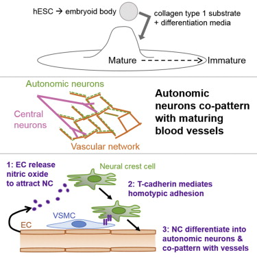
Clues to how human neurovascular unit forms Crucial functions we depend on but don't consciously think about — things like heart rate, blood flow, breathing and digestion — are regulated by our neurovascular unit (1) which is made up of blood vessels and smooth muscles. But how they work together to coordinate functions is not yet understood. Using human embryonic stem cells, researchers at University of California, San Diego School of Medicine and Moores Cancer Center and Sanford-Burnham Medical Research Institute created a model that allows them to track cellular behavior during the earliest stages of human development in real-time. The model reveals, for the first time, how autonomic neurons and blood vessels come together to form the neurovascular unit. The study is published May 21 by Stem Cell Reports.
The neurovascular unit is made up of three cells types: 1. endothelial cells, which form the blood vessel (vascular) tube; 2. smooth muscle cells, which cover the endothelial tube and control vascular tone; and 3. autonomic neurons, which influence smooth muscle's ability to contract and maintain vascular tone. The study revealed that separate signals produced by endothelial cells and smooth muscle cells are required for embryonic cells to differentiate into autonomic neurons. Researchers discovered that endothelial cells secrete nitric oxide, while smooth muscle cells use the protein T-cadherin to interact with neural crest cells, those specialized embryonic cells that give rise to portions of the nervous system and other organs.
In addition to improving the odds that science will one day generate artificial organs from stem cells, this new insight has implications for rare inherited conditions such as neurofibro-matosis, tuberous sclerosis and Hirschsprung's disease.
(1) In the brain, pericytes help sustain the blood–brain barrier as well as several other homeostatic and hemostatic functions of the brain. These cells are also a key component of the neurovascular unit, which includes endothelial cells, astrocytes, and neurons. Pericytes regulate capillary blood flow, the clearance and phagocytosis of cellular debris, and the permeability of the blood–brain barrier. Pericytes stabilize and monitor the maturation of endothelial cells by means of direct communication between the cell membrane as well as through paracrine signaling. A deficiency of pericytes in the central nervous system can cause the blood–brain barrier to break down. Wikipedia Abstract Co-authors include Lisette M. Acevedo, Jeffrey N. Lindquist, UC San Diego and Sanford-Burnham; Breda M. Walsh, Peik Sia, UC San Diego; Flavio Cimadamore, Connie Chen, Martin Denzel, Cameron D. Pernia, Barbara Ranscht, and Alexey Terskikh, Sanford-Burnham. This research was funded, in part, by the National Institutes of Health (grants K01CA148897 and P20GM075059) and California Institute for Regenerative Medicine (grants CIRM-CL1-00511-1 and CIRM-RB3-02098).
|
||||||||||||||||||||||||||||


 co-patterning with blood vessels (red).jpg)