|
|
|
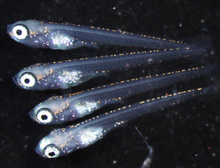
Top: Baby medaka fish born of eggs and sperm from a female fish with nonfuntioning foxl3 gene.
Image Credit: National Institute for Basic Biology
|
|
|
A genetic switch is discovered that determines the fate of germ cells in the ovary of a little fish called the Japanese rice fish or medaka. It turns out that female medaka can produce sperm OR eggs in their ovaries.
For the first time, Japanese researchers have found a genetic switch that determines, in vertebrates, whether germ cells become sperm or eggs.
The gene is named foxl3 and has been identified in the medaka where it was found primarily active in a female's germ cells to prevent them from becoming sperm cells instead of egg cells. Without the functioning of the fox13 gene, sperm AND eggs are produced in the ovaries of medaka females. The sperm are normal and there are confirmed normal offspring from females producing both sperm and eggs. When the scientists inactivated the gene in female fish, the germ cells turned into sperm in the medaka's ovaries rather than eggs.
The work was published in the journal Science, June 11, 2015.
Drs. Toshiya Nishimura, Minoru Tanaka and their colleagues, in collaboration with Drs. Satoru Kobayashi, Mikita Suyama and Yasuyuki Ohkawa, revealed that the foxl3 gene works in the germ cells of females "to suppress eggs from differentiation into sperm."
In females lacking functional foxl3 genes, the small fish appears totally female, however a large number of sperm are formed in the ovaries — along with a small number of eggs — both at the same time. In females lacking a functioning foxl3 gene to suppress sperm production, sperm can be 'made' in a shorter period of time than in normal (wild-type) males.
Researchers are already looking for applications for this discovery in the aquaculture industry.
Previously,
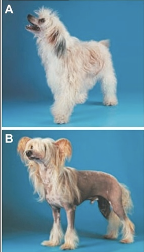
the fox13 gene identified as a switch in the production of hairlessness in dogs. Publshed in Science September 12, 2008, the research established that the Mexican and Peruvian hairless and Chinese crested dogs are all characterized by missing hair and teeth, called canine ectodermal dysplasia (CED). The CED mutation was mapped to an interval on chromosome 17. The associated interval contains a previously uncharacterized member of the forkhead box transcription factor family (FOXI3), which is specifically expressed in developing hair and teeth. So, FOXI3 is seen as a regulator of development in the fetal ectoderm layer in very early embryos.
"In spite of the environment surrounding the germ cells being female, the fact that functional sperm have been made surprised me greatly. That this sexual switch is present in the germ cells — and is independent of the body's sex — is an entirely new finding," Dr. Nishimura said.
"While germ cells can become either sperm or eggs, nobody knew that in vertebrates, the germ cells have a switch mechanism to decide their fate. Our result indicates that once the decision is made the germ cells have the ability to go all the way to the end [of germ cell specialization]."
Minoru Tanaka PhD, Associate Professor, National Institute for Basic Biology, National Institutes of Natural Sciences, Japan
Foxl3 is a germ cell-intrinsic factor involved in sperm-egg fate decision in medaka
Abstract
Sex determination is an essential step in the commitment of a germ cell to a sperm or egg. However, the intrinsic factors that determine the sexual fate of vertebrate germ cells are unknown. Here we show that foxl3, which is expressed in germ cells but not somatic cells in the gonad, is involved in sperm–egg fate decision in medaka fish. Adult XX medaka with disrupted foxl3 developed functional sperm in the expanded germinal epithelium of a histologically functional ovary. In chimeric medaka, mutant germ cells initiated spermatogenesis in female wild-type gonad. These results indicate that a germ cell–intrinsic cue for the sperm–egg fate decision is present in medaka and that spermatogenesis can proceed in a female gonadal environment.
Authors
Toshiya Nishimura, Tetsuya Sato, Yasuhiro Yamamoto, Ikuko Watakabe, Yasuyuki Ohkawa, Mikita Suyama, Satoru Kobayashi2, Minoru Tanaka
Universities involved were: the National Institute for Basic Biology, National Institutes of Natural Sciences in Japan, the Okazaki Institute for Integrative Bioscience, and Kyushu University.
A Mutation in Hairless Dogs Implicates FOXI3 in Ectodermal Development
Abstract
Mexican and Peruvian hairless dogs and Chinese crested dogs are characterized by missing hair and teeth, a phenotype termed canine ectodermal dysplasia (CED). CED is inherited as a monogenic autosomal semidominant trait. With genomewide association analysis we mapped the CED mutation to a 102–kilo–base pair interval on chromosome 17. The associated interval contains a previously uncharacterized member of the forkhead box transcription factor family (FOXI3), which is specifically expressed in developing hair and teeth. Mutation analysis revealed a frameshift mutation within the FOXI3 coding sequence in hairless dogs. Thus, we have identified FOXI3 as a regulator of ectodermal development.
Return to top of page
|


 .
.