|
|
|
Home | Pregnancy Timeline | News Alerts |News Archive Jul 7, 2015
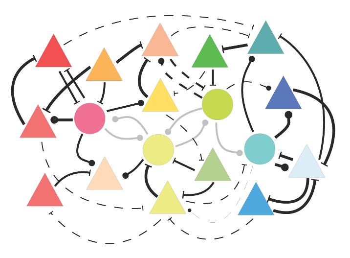
The connections between nerve cells that react to similar stimuli are strengthened
as they
gain visual experience (thick lines) — while other connections are weakened (thin lines).
Image Credit:Stefan Rotter / Bernstein Center Freiburg, 2015 |
|
|
|
|
|
Fine tuning the brain
When newborn babies open their eyes for the first time, they already possess nerve cells specialized to receive stimuli and send it to the visual cortex of their brains - but how do these visual nerve cells link with each other?
How do neural networks which react in a particular way to a particular stimuli, develop over the course of time? In order to better understand the steps and explain the complicated processes of neural organization involved, an international team of researchers developed a computer model that simulates the process.
The study was led by Stefan Rotter, Professor at the Bernstein Center Freiburg (BCF) and Cluster of Excellence BrainLinks — BrainTools at the University of Freiburg, Germany, and Claudia Clopath PhD, Assistant Professor [Lecturer] from the Imperial College London, England. Their work is published in the journals 1.PLOS Computational Biology and 2.PLOS ONE.
"Our model enabled us for the first time ever to combine typical features of biological neural networks in animals and humans via a computer simulation. Neural networks harness feedback to make nerve cells in the visual system more efficient at detecting unique features. These networks can precisely coordinate points of contact between the synapse responses which are inherent in learning."
Sadra Sadeh PhD, neuroscientist, Bernstein Center, Freiburg, Germany.
It is difficult to combine these properties in computer models, because it can easily lead to an explosion of activity in the network - similar to an epileptic fit. To keep the activity in the network stable, the researchers integrated inhibitory synapses into the learning process to control excitation in the network.
Researchers can now use the computer model to simulate various developmental processes in the brain's visual cortex.
Among other things, it will allow scientists to determine how connections between nerve cells change when, for the first time, visual stimuli is received through both eyes following birth. Such processes play a role in early-childhood visual disorders like congenital strabismus — or squinting. "In the long term, the model could even enable us to develop better strategies for treating such illnesses," adds Rotter.
But why do neural networks change their structures in the course of a visual experience — if nerve cells are already specialized the moment our eyes first open? The team found an answer to this question in a parallel study.
"In a simulation comparing inexperienced and fully developed nerve cell networks, we found that fully developed networks strengthen a stimulus to carry more information — by choosing neural connections with the same function."
Stefan Rotter PhD, Bernstein Center Freiburg, and Cluster of Excellence BrainLinks — BrainTools, University of Freiburg, Germany.
Therefore, while newborns do indeed have the capacity to process all stimuli when they first open their eyes, their perception is greatly improved through fine tuning nerve cell connections.
1. Emergence of Functional Specificity in Balanced Networks with Synaptic Plasticity
Abstract
In rodent visual cortex, synaptic connections between orientation-selective neurons are unspecific at the time of eye opening, and become to some degree functionally specific only later during development. An explanation for this two-stage process was proposed in terms of Hebbian plasticity based on visual experience that would eventually enhance connections between neurons with similar response features. For this to work, however, two conditions must be satisfied: First, orientation selective neuronal responses must exist before specific recurrent synaptic connections can be established. Second, Hebbian learning must be compatible with the recurrent network dynamics contributing to orientation selectivity, and the resulting specific connectivity must remain stable for unspecific background activity. Previous studies have mainly focused on very simple models, where the receptive fields of neurons were essentially determined by feedforward mechanisms, and where the recurrent network was small, lacking the complex recurrent dynamics of large-scale networks of excitatory and inhibitory neurons. Here we studied the emergence of functionally specific connectivity in large-scale recurrent networks with synaptic plasticity. Our results show that balanced random networks, which already exhibit highly selective responses at eye opening, can develop feature-specific connectivity if appropriate rules of synaptic plasticity are invoked within and between excitatory and inhibitory populations. If these conditions are met, the initial orientation selectivity guides the process of Hebbian learning and, as a result, functionally specific and a surplus of bidirectional connections emerge. Our results thus demonstrate the cooperation of synaptic plasticity and recurrent dynamics in large-scale functional networks with realistic receptive fields, highlight the role of inhibition as a critical element in this process, and paves the road for further computational studies of sensory processing in neocortical network models equipped with synaptic plasticity.
Author Summary
In primary visual cortex of mammals, neurons are selective to the orientation of contrast edges. In some species, as cats and monkeys, neurons preferring similar orientations are adjacent on the cortical surface, leading to smooth orientation maps. In rodents, in contrast, such spatial orientation maps do not exist, and neurons of different specificities are mixed in a salt-and-pepper fashion. During development, however, a “functional” map of orientation selectivity emerges, where connections between neurons of similar preferred orientations are selectively enhanced. Here we show how such feature-specific connectivity can arise in realistic neocortical networks of excitatory and inhibitory neurons. Our results demonstrate how recurrent dynamics can work in cooperation with synaptic plasticity to form networks where neurons preferring similar stimulus features connect more strongly together. Such networks, in turn, are known to enhance the specificity of neuronal responses to a stimulus. Our study thus reveals how self-organizing connectivity in neuronal networks enable them to achieve new or enhanced functions, and it underlines the essential role of recurrent inhibition and plasticity in this process.
2. Processing of Feature Selectivity in Cortical Networks with Specific Connectivity
Abstract
Although non-specific at the onset of eye opening, networks in rodent visual cortex attain a non-random structure after eye opening, with a specific bias for connections between neurons of similar preferred orientations. As orientation selectivity is already present at eye opening, it remains unclear how this specificity in network wiring contributes to feature selectivity. Using large-scale inhibition-dominated spiking networks as a model, we show that feature-specific connectivity leads to a linear amplification of feedforward tuning, consistent with recent electrophysiological single-neuron recordings in rodent neocortex. Our results show that optimal amplification is achieved at an intermediate regime of specific connectivity. In this configuration a moderate increase of pairwise correlations is observed, consistent with recent experimental findings. Furthermore, we observed that feature-specific connectivity leads to the emergence of orientation-selective reverberating activity, and entails pattern completion in network responses. Our theoretical analysis provides a mechanistic understanding of subnetworks’ responses to visual stimuli, and casts light on the regime of operation of sensory cortices in the presence of specific connectivity.
The Bernstein Center Freiburg is part of the National Bernstein Network Computational Neuroscience in Germany. With this funding initiative, the German Federal Ministry of Education and Research (BMBF) has supported the new discipline of Computational Neuroscience since 2004 with over 180 million Euros. The network is named after the German physiologist Julius Bernstein (1835-1917).
Return to top of page
|


