Welcome to The Visible Embryo
  o
Home-- -History-- -Bibliography- -Pregnancy Timeline- --Prescription Drugs in Pregnancy- -- Pregnancy Calculator- --Female Reproductive System- -Contact
 

Welcome to The Visible Embryo, a comprehensive educational resource on human development from conception to birth.

The Visible Embryo provides visual references for changes in fetal development throughout pregnancy and can be navigated via fetal development or maternal changes.

The National Institutes of Child Health and Human Development awarded Phase I and Phase II Small Business Innovative Research Grants to develop The Visible Embryo. Initally designed to evaluate the internet as a teaching tool for first year medical students, The Visible Embryo is linked to over 600 educational institutions and is viewed by more than one million visitors each month.

Today, The Visible Embryo is linked to over 600 educational institutions and is viewed by more than 1 million visitors each month. The field of early embryology has grown to include the identification of the stem cell as not only critical to organogenesis in the embryo, but equally critical to organ function and repair in the adult human. The identification and understanding of genetic malfunction, inflammatory responses, and the progression in chronic disease, begins with a grounding in primary cellular and systemic functions manifested in the study of the early embryo.

WHO International Clinical Trials Registry Platform


The World Health Organization (WHO) has created a new Web site to help researchers, doctors and patients obtain reliable information on high-quality clinical trials. Now you can go to one website and search all registers to identify clinical trial research underway around the world!


Home

History

Bibliography

Pregnancy Timeline

Prescription Drug Effects on Pregnancy

Pregnancy Calculator

Female Reproductive System

Contact The Visible Embryo

News Alerts Archive

Disclaimer: The Visible Embryo web site is provided for your general information only. The information contained on this site should not be treated as a substitute for medical, legal or other professional advice. Neither is The Visible Embryo responsible or liable for the contents of any websites of third parties which are listed on this site.
Content protected under a Creative Commons License.

No dirivative works may be made or used for commercial purposes.


Pregnancy Timeline by SemestersDevelopmental TimelineFertilizationFirst TrimesterSecond TrimesterThird TrimesterFirst Thin Layer of Skin AppearsEnd of Embryonic PeriodEnd of Embryonic PeriodFemale Reproductive SystemBeginning Cerebral HemispheresA Four Chambered HeartFirst Detectable Brain WavesThe Appearance of SomitesBasic Brain Structure in PlaceHeartbeat can be detectedHeartbeat can be detectedFinger and toe prints appearFinger and toe prints appearFetal sexual organs visibleBrown fat surrounds lymphatic systemBone marrow starts making blood cellsBone marrow starts making blood cellsInner Ear Bones HardenSensory brain waves begin to activateSensory brain waves begin to activateFetal liver is producing blood cellsBrain convolutions beginBrain convolutions beginImmune system beginningWhite fat begins to be madeHead may position into pelvisWhite fat begins to be madePeriod of rapid brain growthFull TermHead may position into pelvisImmune system beginningLungs begin to produce surfactant
CLICK ON weeks 0 - 40 and follow along every 2 weeks of fetal development


Google

Home | Pregnancy Timeline | News Alerts |News Archive Jul 8, 2015


Autism mutations may influence brain size through
RHOA pathway during fetal brain development.
Image Credit: UC San Diego School of Medicine


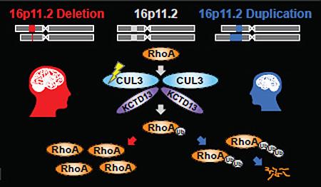
Autism affects neural pathways and brain shape

Scientists have found that mutations that cause autism in children are connected to a neural pathway regulating brain shape during fetal development.


Researchers studying a set of well-known autism mutations called copy number variants or CNVs, found when and where these genes are expressed (turned on) in brain development is significant. "One surprising thing that we immediately observed was that different CNVs seemed to be turned on in different developmental periods," said Lilia Iakoucheva, PhD, assistant professor in the Department of Psychiatry, at the University of California, San Diego School of Medicine, and leader of the research.

Her work is published in Neuron.

Specifically, the team noted that one copy number variation (CNV) located in a genome region known as 16p11.2, contained genes that turn on (become active) during the later part of the mid-fetal period. Ultimately, they identified a network of genes showing a similar pattern of activation including KCTD13 within the same region 16p11.2  — and CUL3, a gene from a different chromosome but also mutated in children with autism.


"The most exciting moment for us was when we realized that proteins encoded by these genes form a complex that regulates levels of a third protein, RhoA. Suddenly, everything came together and made sense," as Rho proteins play critical roles in neuronal migration and brain shape.

Lilia Iakoucheva, PhD, Assistant Professor, Department of Psychiatry, University of California, San Diego School of Medicine, and research leader.


Further experiments confirmed that CUL3 mutations disrupt interaction with the KCTD13 gene, which suggests that the 16p11.2 copy number variation (CNV) and CUL3 mutation could be acting via the same RhoA pathway. RhoA levels influence head and body size in zebrafish, a model animal used by geneticists to investigate gene function. Children with 16p11.2 copy number variations (CNVs) also have enlarged or decreased head sizes and suffer from obesity or are underweight.

"Our model fits perfectly with what we observe in patients," adds Guan Ning Lin, PhD, a fellow in Iakoucheva's laboratory and co-first author along with Roser Corominas, PhD.


Interestingly, the RhoA pathway has recently been implicated in a rare form of autism called Timothy syndrome, caused by the CNV mutation on a completely different gene.

"The fact that three different types of mutations may act via the same pathway is remarkable. My hope is that we may be able to target it therapeutically.

"If we can discover the precise mechanism and develop targeted treatments for a handful of children, or even for a single child with autism, I would be happy."


Lilia Iakoucheva, PhD


Iakoucheva and colleagues are planning to test inhibitors of the RhoA pathway using a stem cell model of autism.

Abstract
Highlights
•Rare high-risk CNVs for psychiatric disorders have unique spatiotemporal signatures
•Dynamic 16p11.2 protein interaction network reveals changes during brain development
•The late mid-fetal period is critical for establishing 16p11.2 network connectivity
•KCTD13-Cul3-RhoA pathway may be dysregulated by gene-damaging mutations

Summary
The psychiatric disorders autism and schizophrenia have a strong genetic component, and copy number variants (CNVs) are firmly implicated. Recurrent deletions and duplications of chromosome 16p11.2 confer a high risk for both diseases, but the pathways disrupted by this CNV are poorly defined. Here we investigate the dynamics of the 16p11.2 network by integrating physical interactions of 16p11.2 proteins with spatiotemporal gene expression from the developing human brain. We observe profound changes in protein interaction networks throughout different stages of brain development and/or in different brain regions. We identify the late mid-fetal period of cortical development as most critical for establishing the connectivity of 16p11.2 proteins with their co-expressed partners. Furthermore, our results suggest that the regulation of the KCTD13-Cul3-RhoA pathway in layer 4 of the inner cortical plate is crucial for controlling brain size and connectivity and that its dysregulation by de novo mutations may be a potential determinant of 16p11.2 CNV deletion and duplication phenotypes.

Co-authors include Xinping Yang, David E. Hill and Marc Vidal, Dana-Farber Cancer Institute; Irma Lemmens and Jan Tavernier, Ghent University, Belgium; and Jonathan Sebat, Beyster Center for Genomics of Psychiatric Diseases and UCSD.

This research was funded, in part, by National Institutes of Health (grants R01MH091350, R01HD065288, R21MH104766 and R01MH105524).

Return to top of page