|
|
|
Home | Pregnancy Timeline | News Alerts |News Archive Jul 22, 2015
Small molecules such as sialic acid direct cell
communication and can profoundly affect behavior. |
|
|
|
|
|
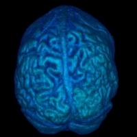
Sialic acid is key to unlocking brain disorders
Research in mice may help us understand how small changes in the way sialic acid attaches to the surface of a neuron can affect motor skills and learning, or cause hyperactivity.
A new report published in the July 2015 issue of The FASEB Journal suggests that a common molecule, sialic acid, found in higher animals including humans, ultimately affects brain structure. Sialic may play a significant role in how brain cells communicate, possibly shedding light on the underlying causes of a number of brain disorders.
Babies are born with a complete number of brain neurons or nerve cells, but synapse connections between neurons continue to expand throughout life. As the human brain has 2 – 4 times more sialic acid than found in other mammal brains, it suggests an integral role for it in brain development and learning, so it is important that a child has an adequate supply.
"Sialic acid is part of the molecular language that cells use to communicate between themselves. As we learn that language, we can use the knowledge to better understand disease and perhaps to thoughtfully intervene."
Ronald L. Schnaar PhD, senior researcher, the Departments of Pharmacology and Neuroscience, Johns Hopkins University School of Medicine, Baltimore, Maryland.
To make their discovery, Schnaar and colleagues mutated mouse genes responsible for sialic acid attachment, then compared the resulting brain structures, motor function, activity, and learning in those mice to normal mice. What they found is that mice with altered sialic acid attachment had significant neurological problems compared to normal mice.
Gerald Weissmann, M.D., Editor-in-Chief of The FASEB Journal: "The molecular codes that control the human brain are as yet poorly worked out. This report shows how small molecules such as sialic acid direct cell communication and if interrupted, can profoundly affect behavior. With this information, researchers have new ways to work out the mechanisms that determine hyperactivity and other brain disorders."
Abstract
Every cell expresses a molecularly diverse surface glycan coat (glycocalyx) comprising its interface with its cellular environment. In vertebrates, the terminal sugars of the glycocalyx are often sialic acids, 9-carbon backbone anionic sugars implicated in intermolecular and intercellular interactions. The vertebrate brain is particularly enriched in sialic acid-containing glycolipids termed gangliosides. Human congenital disorders of ganglioside biosynthesis result in paraplegia, epilepsy, and intellectual disability. To better understand sialoglycan functions in the nervous system, we studied brain anatomy, histology, biochemistry, and behavior in mice with engineered mutations in St3gal2 and St3gal3, sialyltransferase genes responsible for terminal sialylation of gangliosides and some glycoproteins. St3gal2/3 double-null mice displayed dysmyelination marked by a 40% reduction in major myelin proteins, 30% fewer myelinated axons, a 33% decrease in myelin thickness, and molecular disruptions at nodes of Ranvier. In part, these changes may be due to dysregulation of ganglioside-mediated oligodendroglial precursor cell proliferation. Neuronal markers were also reduced up to 40%, and hippocampal neurons had smaller dendritic arbors. Young adult St3gal2/3 double-null mice displayed impaired motor coordination, disturbed gait, and profound cognitive disability. Comparisons among sialyltransferase mutant mice provide insights into the functional roles of brain gangliosides and sialoglycoproteins consistent with related human congenital disorders.—Yoo, S.-W., Motari, M. G., Susuki, K., Prendergast, J., Mountney, A., Hurtado, A., Schnaar, R. L. Sialylation regulates brain structure and function.
Details: Seung-Wan Yoo, Mary G. Motari, Keiichiro Susuki, Jillian Prendergast, Andrea Mountney, Andres Hurtado, and Ronald L. Schnaar. Sialylation regulates brain structure and function. FASEB J. July 2015 29:3040-3053; doi:10.1096/fj.15-270983 ; http://www.fasebj.org/content/29/7/3040.abstract
FASEB is composed of 27 societies with more than 125,000 members, making it the largest coalition of biomedical research associations in the United States. Our mission is to advance health and welfare by promoting progress and education in biological and biomedical sciences through service to our member societies and collaborative advocacy.
Return to top of page
|


