Welcome to The Visible Embryo
    o >
Home-- -History-- -Bibliography- -Pregnancy Timeline- --Prescription Drugs in Pregnancy- -- Pregnancy Calculator- --Female Reproductive System- -Contact
   
Welcome to The Visible Embryo, a comprehensive educational resource on human development from conception to birth.

The Visible Embryo provides visual references for changes in fetal development throughout pregnancy and can be navigated via fetal development or maternal changes.

The National Institutes of Child Health and Human Development awarded Phase I and Phase II Small Business Innovative Research Grants to develop The Visible Embryo. Initally designed to evaluate the internet as a teaching tool for first year medical students, The Visible Embryo is linked to over 600 educational institutions and is viewed by more than one million visitors each month.

Today, The Visible Embryo is linked to over 600 educational institutions and is viewed by more than 1 million visitors each month. The field of early embryology has grown to include the identification of the stem cell as not only critical to organogenesis in the embryo, but equally critical to organ function and repair in the adult human. Identification and understanding of genetic malfunction, inflammatory responses, and the progression in chronic disease, begins with a grounding in primary cellular and systemic functions manifested in the study of the early embryo.

WHO International Clinical Trials Registry Platform

The World Health Organization (WHO) has created a new Web site to help researchers, doctors and patients obtain reliable information on high-quality clinical trials. Now you can go to one website and search all registers to identify clinical trial research underway around the world!


Home

History

Bibliography

Pregnancy Timeline

Prescription Drug Effects on Pregnancy

Pregnancy Calculator

Female Reproductive System

Contact The Visible Embryo

News Alerts Archive

Disclaimer: The Visible Embryo web site is provided for your general information only. The information contained on this site should not be treated as a substitute for medical, legal or other professional advice. Neither is The Visible Embryo responsible or liable for the contents of any websites of third parties which are listed on this site.
Content protected under a Creative Commons License.

No dirivative works may be made or used for commercial purposes.


Pregnancy Timeline by SemestersFetal liver is producing blood cellsHead may position into pelvisBrain convolutions beginFull TermWhite fat begins to be madeWhite fat begins to be madeHead may position into pelvisImmune system beginningImmune system beginningPeriod of rapid brain growthBrain convolutions beginLungs begin to produce surfactantSensory brain waves begin to activateSensory brain waves begin to activateInner Ear Bones HardenBone marrow starts making blood cellsBone marrow starts making blood cellsBrown fat surrounds lymphatic systemFetal sexual organs visibleFinger and toe prints appearFinger and toe prints appearHeartbeat can be detectedHeartbeat can be detectedBasic Brain Structure in PlaceThe Appearance of SomitesFirst Detectable Brain WavesA Four Chambered HeartBeginning Cerebral HemispheresFemale Reproductive SystemEnd of Embryonic PeriodEnd of Embryonic PeriodFirst Thin Layer of Skin AppearsThird TrimesterSecond TrimesterFirst TrimesterFertilizationDevelopmental Timeline
CLICK ON weeks 0 - 40 and follow along every 2 weeks of fetal development


Google Search artcles published since 2007


Home | Pregnancy Timeline | News Alerts |News Archive Jul 28, 2015 
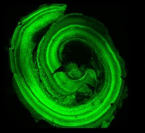
Studying mice, scientists have identified two signaling molecules required for the development
of the cochlea in the inner ear. A normal mouse cochlea shows a characteristic spiral shape
(above). Without both signals, the embryo cannot produce enough cells, resulting in a
shortened cochlear duct and impaired hearing.
Image Credit: Sung-Ho Huh


Reverse hearing loss?

Unlike birds and amphibians, mammals can't recover lost hearing. In people, the cells of the inner ear responsible for detecting sound and transmitting those signals to the brain form during early weeks of development. And unlike some animals, can't be replaced if lost illness, injury or aging.


In people, the cells of the inner ear responsible for detecting sound and transmitting its signals to the brain, form during the early stages of human development. And, they can't be replaced if lost to illness, injury or aging.

Using mice as their model, scientists at Washington University School of Medicine in St. Louis have identified two signaling molecules required for development of the cochlea in the inner ear. They found without both signals, the embryo cannot produce enough cells to make the cochlea. This results in a shortened cochlear duct and impaired hearing.

The study is available in eLife and contributes to our understanding of inner ear development.

"To eventually be able to restore hearing, we would like to regenerate the sensory hair cells of the cochlea," said senior author David M. Ornitz, MD, PhD, the Alumni Endowed Professor of Developmental Biology. "If the inner ear in birds and fish is damaged, for example, cells are naturally converted back into progenitor cells, capable of replacing the sensory cells. But mammals are more complex — with a better sense of hearing over a wider range of sounds. It is thought that in exchange for better hearing, we have lost the ability to regenerate sensory hair cells."


In the new study, Ornitz and his colleagues showed that proper mouse inner ear development depends on two molecules called FGF9 and FGF20.


These two molecules begin signalling in the inner ear, about day 11 of the typical 20-days of mouse embryo development. Over the next two to three days, they alert progenitor cells to multiply. By day 14, progenitor cells stop multiplying and begin to differentiate into functional adult sensory cells. At this point, the cell population making up the adult ear is largely complete.


"In mammals, mice and people included, the number of sensory progenitor cells is fixed. That number is determined by cell division or cell death in early stages of development. In mice, this is between embryonic days 11 and 14. Once that developmental window is closed, the number of cells you have is all you get. There is no compensating for low numbers."

Sung-Ho Huh PhD, first author, professor of developmental biology.


The hair cells of the inner ear pick up sound vibrations and transmit those signals to the brain. Hearing loss occurs when these hair cells are damaged, most often by loud noise, some types of medications and just aging itself.  FGF9 and FGF20 send signals to receptors located in developing sensory progenitor cells that stimulate their growth — this activates a feedback loop directing development of the cochlea. Ornitz and Huh in their future research will focus on identifying all of the molecules involved in this feedback mechanism.


"We have discovered that an FGF signal is instructive in forming the cochlea. These FGF signals tell the surrounding tissue to make a factor — we don't know yet what that factor is — but we know it regulates progenitor cell growth. And being able to grow progenitor cells, or instruct cells that can become progenitor cells to grow, is one key to restoring hearing."

David M. Ornitz, MD, PhD, the Alumni Endowed Professor of Developmental Biology, Washington University School of Medicine, St. Louis, Missouri.


Abstract
The sensory and supporting cells (SCs) of the organ of Corti are derived from a limited number of progenitors. The mechanisms that regulate the number of sensory progenitors are not known. Here, we show that Fibroblast Growth Factors (FGF) 9 and 20, which are expressed in the non-sensory (Fgf9) and sensory (Fgf20) epithelium during otic development, regulate the number of cochlear progenitors. We further demonstrate that Fgf receptor (Fgfr) 1 signaling within the developing sensory epithelium is required for the differentiation of outer hair cells and SCs, while mesenchymal FGFRs regulate the size of the sensory progenitor population and the overall cochlear length. In addition, ectopic FGFR activation in mesenchyme was sufficient to increase sensory progenitor proliferation and cochlear length. These data define a feedback mechanism, originating from epithelial FGF ligands and mediated through periotic mesenchyme that controls the number of sensory progenitors and the length of the cochlea.

This work was supported by the Action on Hearing Loss Foundation; the Office of Naval Research, grant number N000141211025; the March of Dimes Foundation; the Hearing Health Foundation; and the National Institutes of Health (NIH), grant numbers K99 DC012825, P30 DC004665, P30 DK052574 and P30 AR057235.

Huh SH, Warchol ME, Ornitz DM. Cochlear progenitor number is controlled through mesenchymal FGF receptor signaling. eLife. Online April 27, 2015.

Washington University School of Medicine's 2,100 employed and volunteer faculty physicians also are the medical staff of Barnes-Jewish and St. Louis Children's hospitals. The School of Medicine is one of the leading medical research, teaching and patient-care institutions in the nation, currently ranked sixth in the nation by U.S. News & World Report. Through its affiliations with Barnes-Jewish and St. Louis Children's hospitals, the School of Medicine is linked to BJC HealthCare.

Additional reading: Gene modulation method may provide insight on regrowing inner-ear sensory hair cells

Return to top of page