Welcome to The Visible Embryo
  o
 
The Visible Embryo Home
   
Google  
Home--- -History-----Bibliography-----Pregnancy Timeline-----Prescription Drugs in Pregnancy---- Pregnancy Calculator----Female Reproductive System----News Alerts----Contact
 
 
Welcome to The Visible Embryo, a comprehensive educational resource on human development from conception to birth.

The Visible Embryo provides visual references for changes in fetal development throughout pregnancy and can be navigated via fetal development or maternal changes.

The National Institutes of Child Health and Human Development awarded Phase I and Phase II Small Business Innovative Research Grants to develop The Visible Embryo. Initally designed to evaluate the internet as a teaching tool for first year medical students, The Visible Embryo is linked to over 600 educational institutions and is viewed by more than one million visitors each month.

Today, The Visible Embryo is linked to over 600 educational institutions and is viewed by more than 1 million visitors each month. The field of early embryology has grown to include the identification of the stem cell as not only critical to organogenesis in the embryo, but equally critical to organ function and repair in the adult human. The identification and understanding of genetic malfunction, inflammatory responses, and the progression in chronic disease, begins with a grounding in primary cellular and systemic functions manifested in the study of the early embryo.

WHO International Clinical Trials Registry Platform


The World Health Organization (WHO) has created a new Web site to help researchers, doctors and patients obtain reliable information on high-quality clinical trials. Now you can go to one website and search all registers to identify clinical trial research underway around the world!


Home

History

Bibliography

Pregnancy Timeline

Prescription Drug Effects on Pregnancy

Pregnancy Calculator

Female Reproductive System

Contact The Visible Embryo

News Alerts Archive

Disclaimer: The Visible Embryo web site is provided for your general information only. The information contained on this site should not be treated as a substitute for medical, legal or other professional advice. Neither is The Visible Embryo responsible or liable for the contents of any websites of third parties which are listed on this site.
Content protected under a Creative Commons License.

No dirivative works may be made or used for commercial purposes.

 
Pregnancy Timeline by SemestersDevelopmental TimelineFertilizationFirst TrimesterSecond TrimesterThird TrimesterFirst Thin Layer of Skin AppearsEnd of Embryonic PeriodEnd of Embryonic PeriodFemale Reproductive SystemBeginning Cerebral HemispheresA Four Chambered HeartFirst Detectable Brain WavesThe Appearance of SomitesBasic Brain Structure in PlaceHeartbeat can be detectedHeartbeat can be detectedFinger and toe prints appearFinger and toe prints appearFetal sexual organs visibleBrown fat surrounds lymphatic systemBone marrow starts making blood cellsBone marrow starts making blood cellsInner Ear Bones HardenSensory brain waves begin to activateSensory brain waves begin to activateFetal liver is producing blood cellsBrain convolutions beginBrain convolutions beginImmune system beginningWhite fat begins to be madeHead may position into pelvisWhite fat begins to be madePeriod of rapid brain growthFull TermHead may position into pelvisImmune system beginningLungs begin to produce surfactant
CLICK ON weeks 0 - 40 and follow along every 2 weeks of fetal development


  Altered brain growth in autism spectrum disorder

Scripps Research Institute has found that mutations in the autism risk gene — PTEN —
lead to overproduction of the neurons and glia that build the cerebral cortex
(visualized here using a red fluorescent reporter).
Image Credit: The Scripps Research Institute.


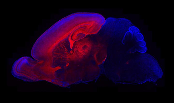
More than half a century after autism was identified by psychiatrist Leo Kanner in 1943, the exact causes of this brain disorder still remain unclear. Now Scripps research has uncovered how mutations in the gene PTEN, mutated in 20% of autism cases, alters early brain development in mice and contributes to macrocephaly or enlarged head.


The study is published in the July 15 issue of The Journal of Neuroscience, and focuses on the gene PTEN (Phosphatase and tensin homolog), which is mutated in around 20 percent of individuals with autism spectrum disorder with macrocephaly (enlarged head).

Autism spectrum disorder is characterized by social deficits, repetitive behaviors and interests, difficulty in communication, as well as cognitive delays in some individuals. It affects approximately one percent of the population with about 80 percent being male.

Scripps Florida biologist Damon Page and team, observed that mutations in the PTEN gene approximately reflects the percentage of a subgroup of individuals with autism spectrum disorder with enlarged heads. These PTEN mutations lead to increases in two key cell types making up the brain — neurons and glial cells. In adult mice, the number of neurons in the brains of PTEN mutants is virtually the same as in normal mice. However, glia cells — which support neurons — are overrepresented.


"In the adult brain, excess glia are a primary cause of a change in brain size. This raises the intriguing possibility that these excess glia may in fact contribute to abnormal development and thus function in brain circuitry, when PTEN is mutated."

Damon Page PhD, Department of Neuroscience, The Scripps Research Institute, Jupiter, Florida


Brain overgrowth is a dynamic process. The greatest increase in size occurrs at birth and then again in adulthood. The least overgrowth is in the early juvenile. The team observed that this abnormal growth pattern appears to be sparked by amplification of neuron development. Before birth, neurons are trimmed by a process of programmed cell death (called apoptosis), and glia cells development follows after neurons.

"Apoptosis is a natural phenomenon that removes unnecessary neurons during normal brain development," says Research Associate Youjun Chen, the first author of the study and a member of the Page laboratory. "We find it very striking that in brains of PTEN mutant mice, the presence of excess neurons is corrected by excessive apoptosis. After that, excess glia are then made. In adulthood, the number of glial cells continues to increase by more than 20 percent in our [mouse] models."

The scientists traced these effects back to an increase in signals from a molecule known as β-Catenin (beta Catenin).


"PTEN and β-catenin are two important molecules that control growth in the developing brain in both mice and humans. They work together to regulate brain growth by controlling the number and types of cells being produced... this suggests that an imbalance in this relationship may contribute to abnormal brain growth in a subset of individuals with autism spectrum disorder."


Interestingly, Page noted that in spite of the profound effects of PTEN mutations on brain growth, the mice are largely able to adapt at the level of behavior, with the important exception of social behavior and a few other behaviors relevant to autism spectrum disorder.

Page: "Our findings across studies indicate that it may be a multiple-hit process. While abnormal growth puts stress on the developing brain, the brain works hard to compensate. How well an individual can adapt to an abnormal pattern of brain growth may shape their outcome in terms of behavior and cognition. The capacity to adapt may, in turn, be influenced by genetic or environmental factors."

Abstract
Abnormal patterns of head and brain growth are a replicated finding in a subset of individuals with autism spectrum disorder (ASD). It is not known whether risk factors associated with ASD and abnormal brain growth (both overgrowth and undergrowth) converge on common biological pathways and cellular mechanisms in the developing brain. Heterozygous mutations in PTEN (PTEN+/−), which encodes a negative regulator of the PI3K-Akt-mTOR pathway, are a risk factor for ASD and macrocephaly. Here we use the developing cerebral cortex of Pten+/− mice to investigate the trajectory of brain overgrowth and underlying cellular mechanisms. We find that overgrowth is detectable from birth to adulthood, is driven by hyperplasia, and coincides with excess neurons at birth and excess glia in adulthood. β-Catenin signaling is elevated in the developing Pten+/− cortex, and a heterozygous mutation in Ctnnb1 (encoding β-catenin), itself a candidate gene for ASD and microcephaly, can suppress Pten+/− cortical overgrowth. Thus, a balance of Pten and β-catenin signaling regulates normal brain growth trajectory by controlling cell number, and imbalance in this relationship can result in abnormal brain growth.

Significance Statement
We report that Pten haploinsufficiency leads to a dynamic trajectory of brain overgrowth during development and altered scaling of neuronal and glial cell populations. β-catenin signaling is elevated in the developing cerebral cortex of Pten haploinsufficient mice, and a heterozygous mutation in β-catenin, itself a candidate gene for ASD and microcephaly, suppresses Pten+/− cortical overgrowth. This leads to the new insight that Pten and β-catenin signaling act in a common pathway to regulate normal brain growth trajectory by controlling cell number, and disruption of this pathway can result in abnormal brain growth.

In addition to Page and Chen, other authors of the study, "Pten Mutations Alter Brain Growth Trajectory and Allocation of Cell Types through Elevated β-Catenin Signaling," are Wen-Chin Huang, Julien Sejourne and Amy E. Clipperton-Allen of TSRI.

This work was supported by the state of Florida, Ms. Nancy Lurie Marks, the Simons Foundation (grant award 360712), an O'Keeffe Neuroscience Scholars Award and the Fraternal Order of Eagles.

Return to top of page