Welcome to The Visible Embryo
  o
 
The Visible Embryo Home
   
Google  
Home--- -History-----Bibliography-----Pregnancy Timeline-----Prescription Drugs in Pregnancy---- Pregnancy Calculator----Female Reproductive System----News Alerts----Contact
 

Welcome to The Visible Embryo, a comprehensive educational resource on human development from conception to birth.

The Visible Embryo provides visual references for changes in fetal development throughout pregnancy and can be navigated via fetal development or maternal changes.

The National Institutes of Child Health and Human Development awarded Phase I and Phase II Small Business Innovative Research Grants to develop The Visible Embryo. Initally designed to evaluate the internet as a teaching tool for first year medical students, The Visible Embryo is linked to over 600 educational institutions and is viewed by more than one million visitors each month.

Today, The Visible Embryo is linked to over 600 educational institutions and is viewed by more than 1 million visitors each month. The field of early embryology has grown to include the identification of the stem cell as not only critical to organogenesis in the embryo, but equally critical to organ function and repair in the adult human. The identification and understanding of genetic malfunction, inflammatory responses, and the progression in chronic disease, begins with a grounding in primary cellular and systemic functions manifested in the study of the early embryo.

WHO International Clinical Trials Registry Platform


The World Health Organization (WHO) has created a new Web site to help researchers, doctors and patients obtain reliable information on high-quality clinical trials. Now you can go to one website and search all registers to identify clinical trial research underway around the world!


Home

History

Bibliography

Pregnancy Timeline

Prescription Drug Effects on Pregnancy

Pregnancy Calculator

Female Reproductive System

Contact The Visible Embryo

News Alerts Archive

Disclaimer: The Visible Embryo web site is provided for your general information only. The information contained on this site should not be treated as a substitute for medical, legal or other professional advice. Neither is The Visible Embryo responsible or liable for the contents of any websites of third parties which are listed on this site.


Content protected under a Creative Commons License.
No dirivative works may be made or used for commercial purposes.


Pregnancy Timeline by SemestersDevelopmental TimelineFertilizationFirst TrimesterSecond TrimesterThird TrimesterFirst Thin Layer of Skin AppearsEnd of Embryonic PeriodEnd of Embryonic PeriodFemale Reproductive SystemBeginning Cerebral HemispheresA Four Chambered HeartFirst Detectable Brain WavesThe Appearance of SomitesBasic Brain Structure in PlaceHeartbeat can be detectedHeartbeat can be detectedFinger and toe prints appearFinger and toe prints appearFetal sexual organs visibleBrown fat surrounds lymphatic systemBone marrow starts making blood cellsBone marrow starts making blood cellsInner Ear Bones HardenSensory brain waves begin to activateSensory brain waves begin to activateFetal liver is producing blood cellsBrain convolutions beginBrain convolutions beginImmune system beginningWhite fat begins to be madeHead may position into pelvisWhite fat begins to be madePeriod of rapid brain growthFull TermHead may position into pelvisImmune system beginningLungs begin to produce surfactant
CLICK ON weeks 0 - 40 and follow along every 2 weeks of fetal development


Fetal Timeline      Maternal Timeline      News     News Archive    Aug 11, 2015 


 2 patients with Prader-Willi Syndrome, exhibiting characteristic facial appearance, with an elongated face, thin upper lip and a prominent nose. a) a 15-year-old male. b) a 41-year-old female.


This painting inspired the initial description of Angelman Syndrome


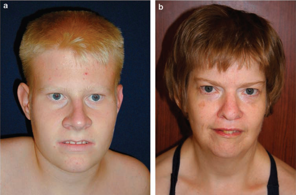
How a single genetic mutation can cause autism

New research shows the precise cell mechanisms that leads to autism disorder, and an existing drug which might help thousands of people with autism.


Last December it was announced that more than 1,000 gene mutations in people with autism had been identified, but it was left unexplained how these particular mutations increased the risk for autism. Now, researchers at the University of North Carolina (UNC) School of Medicine have found how one disabled molecular switch in one gene can lead to the disorder, while duplication or triplication of the same switch can lead to Angelman Syndrome (AS).

AS is a classic example of genomic imprinting. It is caused by deletion or inactivation of genes on the mother's chromosome 15. The father's copy of chromosome 15, which may be normal, is imprinted and therefore silenced. The companion syndrome, Prader-Willi Syndrome, is caused by a similar loss of a father's inherited genes and a mother's genes being imprinted and also silenced.

Ubiquitin-protein ligase E3A, UBE3A, is an enzyme produced by the UBE3A gene, to tag proteins for removal. This is a normal process that maintains healthy cell function by removing damaged or unnecessary proteins. The ligase attaches a small marker, ubiquitin, to unneeded proteins. At that point, structures called proteasomes, recognize and digest any protein so tagged.

UBE3A is switched off when it is tagged with a phosphate molecule. Deleting the function of UBE3A therefore causes cellular chaos. One type of neurodevelopmental disorder that results from this malfunction is Angelman syndrome (AS).  However, duplicating or triplicating UBE3A keeps UBE3A perpetually switched on. As a result, UBE3A becomes hyperactive, creating abnormal dendritic spines all over neurons and results in autism.

The research is published in the journal Cell


"Genetic studies are showing that there will be about 1,000 genes linked to autism. This means you could mutate any one of them and get the disorder. We found how [just] one of these mutations works."

Mark Zylka PhD, Associate Professor, Cell Biology and Physiology, UNC Neuroscience Center, senior author of the paper.


Research was conducted on human cell lines and in mouse models, as Zylka had cells from previous research with an autism-linked UBE3A mutation patient. Sequencing genes from those cell samples - including cells from the child's parents - Zulka found that only the child had hyperactive UBE3A, not the parents. The child's regulatory switch was broken causing UBE3A to be perpetually switched on and ubiquitin not being attached to proteins needing to be destroyed.

"When this child's mutation was introduced into an animal model, we saw all these dendritic spines form on the [mouse's] neurons," explained Zylka, a member of the Carolina Institute for Developmental Disabilities. "Having too many dendritic spines has been linked to autism." Their findings thus pointed to hyperactivation of UBE3A as the likely cause of the child's condition.

Duplication of the 15q chromosome region - which includes UBE3A and several other genes - is one of the most commonly seen genetic alterations in people with autism and is called "Dup15q Syndrome."


As part of their study, Zylka and team found Protein Kinase A (PKA) is the enzyme that tacks the phosphate group onto UBE3A. This finding has therapeutic implications, particularly as drugs exist to control PKA.

Zylka: "We think it may be possible to tamp down UBE3A in Dup15q patients to restore normal levels of enzyme activity in the brain. In fact, we tested known compounds and showed that two of them substantially reduced UBE3A activity in neurons."


One of the drugs, rolipram, previously had been tested in clinical trials treating depression but was discontinued when a patient suffered sudden death by epileptic seizure. In light of this result, Zylka believes retesting must be done with lower doses of rolipram, or any other drug that may increase PKA activity, if only to provide symptom relief for some Dup15q individuals. "The benefits might outweigh the risks," he feels. Such tests would be done with an animal model of Dup15q first.

While the bulk of this project focused on autism, it began by identifying Angelman Syndrome-linked mutations clustered in the same chromosome region where a phosphate group attaches to UBE3A. Zylka's team found that a number of Angelman mutations disrupt the function of UBE3A — and that deleting UBE3A function altogether causes cell chaos. Mutations that could essentially eliminate the UBE3A enzyme in Angelman Syndrome patients, is a discovery which could also help diagnose people with this rare and often misdiagnosed disorder.

Abstract Highlights
•PKA phosphorylates UBE3A at T485 and inhibits UBE3A ubiquitin ligase activity
•Autism-linked UBE3A T485A missense mutation disrupts phosphorylation regulation
•The T485A mutation hyperactivates UBE3A and increases synapse formation in vivo

Summary
Deletion of UBE3A causes the neurodevelopmental disorder Angelman syndrome (AS), while duplication or triplication of UBE3A is linked to autism. These genetic findings suggest that the ubiquitin ligase activity of UBE3A must be tightly maintained to promote normal brain development. Here, we found that protein kinase A (PKA) phosphorylates UBE3A in a region outside of the catalytic domain at residue T485 and inhibits UBE3A activity toward itself and other substrates. A de novo autism-linked missense mutation disrupts this phosphorylation site, causing enhanced UBE3A activity in vitro, enhanced substrate turnover in patient-derived cells, and excessive dendritic spine development in the brain. Our study identifies PKA as an upstream regulator of UBE3A activity and shows that an autism-linked mutation disrupts this phosphorylation control. Moreover, our findings implicate excessive UBE3A activity and the resulting synaptic dysfunction to autism pathogenesis.

Other co-authors included Ben Philpot, PhD, professor of cell biology and physiology, William Snider, MD, director of the UNC Neuroscience Center, and Klaus Hahn, PhD, the Ronald Thurman Distinguished Professor of Pharmacology. Janet Berrios, a graduate student at UNC, and Jason Newbern, PhD, assistant professor in the School of Life Sciences at Arizona State University, are also co-authors.

The National Institutes of Health, the Angelman Syndrome Foundation, The Foundation for Angelman Syndrome Therapeutics, and Autism Speaks funded this work.

Return to top of page