|
|
Welcome to The Visible Embryo, a comprehensive educational resource on human development from conception to birth.
The Visible Embryo provides visual references for changes in fetal development throughout pregnancy and can be navigated via fetal development or maternal changes.
The National Institutes of Child Health and Human Development awarded Phase I and Phase II Small Business Innovative Research Grants to develop The Visible Embryo. Initally designed to evaluate the internet as a teaching tool for first year medical students, The Visible Embryo is linked to over 600 educational institutions and is viewed by more than one million visitors each month.
Today, The Visible Embryo is linked to over 600 educational institutions and is viewed by more than 1 million visitors each month. The field of early embryology has grown to include the identification of the stem cell as not only critical to organogenesis in the embryo, but equally critical to organ function and repair in the adult human. The identification and understanding of genetic malfunction, inflammatory responses, and the progression in chronic disease, begins with a grounding in primary cellular and systemic functions manifested in the study of the early embryo.

The World Health Organization (WHO) has created a new Web site to help researchers, doctors and patients obtain reliable information on high-quality clinical trials. Now you can go to one website and search all registers to identify clinical trial research underway around the world!

|
|
| Disclaimer: The Visible Embryo web site is provided for your general information only. The information contained on this site should not be treated as a substitute for medical, legal or other professional advice. Neither is The Visible Embryo responsible or liable for the contents of any websites of third parties which are listed on this site. |
|
|

Content protected under a Creative Commons License. Commons License. |
|
| No dirivative works may be made or used for commercial purposes. |
|
|
| |
|
|
 
CLICK ON weeks 0 - 40 and follow along every 2 weeks of fetal development
|
|
|
|
Fetal Timeline Maternal Timeline News News Archive Aug 18, 2015
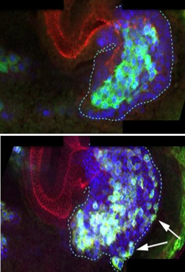
In a developing Drosophila embryo, (TOP) E-Cadherin keep cells together to
facilitate coordinated migration; (BELOW) without E-Cad cells are disorganized.
Image Credit: Jordi Casanova Laboratories
|
|
| |
|
|
|
One protein keeps cells both static and moving!
New research reveals the protein E-Cadherin not only makes a cell "sticky," but is also critical for its mobility.
The protein E-Cadherin (E-Cad) has always been thought of as a kind of adhesive that keeps cells stuck together, thus supporting cells organizing into tissues and organs. Now scientists at the Institute for Research in Biomedicine (IRB Barcelona, Spain) have revealed a new E-Cad function.
Emphasizing the importance of studying early embryonic development, researchers Kyra Campbell and Jordi Casanova, Institut de Biologia Molecular de Barcelona (CSIC), found this new E-Cad phenomenon in the digestive system of embryonic fruit flies. Drosophila melanogaster (fruit fly) is an insect often used by scientists to model cell migration. Investigating how single cells become complicated organisms opens up biological processes critical throughout life, both functional and dysfunctional.
The researchers found E-Cad is crucial for coordinating movement by diverse cell types. This new function may not only help explain how tissues are formed, but why tumors with higher levels of E-Cad have a poorer prognosis, as higher levels of E-Cad may be facilitating tumor cells movement and adhesiveness to other cell types.
The work is published in Nature Communications.
E-Cad facilitates movement by diverse groups of cells. These cell groups produce a range of cell activities due to their different gene sequences. Some cells may divide many times, others may trigger specific hormones, while still others interact only with membranes, and on and on. These cells can move in a coordinated manner to any destination thanks to E-Cad. And once there, they will disemminate into available tissue. Moderate levels of E-Cad have kept the cells bound together, but not immobile.
"Cell migration is a common and necessary process for an embryo and ultimately for the correct function of an adult organism. What has been surprising is the observation that E-Cad is a key component in cell movement. Its role previously was always assumed to be for keeping cells static."
Jordi Casanova PhD, Director, Department of Development and Morphogenesis, Drosophila Lab, IRB Barcelona, The Spanish National Research Council (Consejo Superior de Investigaciones Científicas, CSIC) research professor.
Cell migration is of great biomedical importance. Research into migration helps explain how wounds heal, how inflammation arises and now, how cancer may metastasize.
According to Dr. Casanova, intermediary levels of E-Cad are often associated with aggressive tumors, precisely those capable of metastasising. He adds: "the more we learn about metastases, the more evidence emerges that they [cancers] are formed by groups of cells and not by individual cells. A cell that migrates alone is much easier to eliminate that a group of cells with different functions. Our research in Drosophila is clinically relevant because it offers an explanation for the role which E-Cad may play in tumor metastasis."
Abstract
Collective cell migration is a key process underlying the morphogenesis of many organs as well as tumour invasion, which very often involves heterogeneous cell populations. Here we investigated how such populations can migrate cohesively in the Drosophila posterior midgut, comprised of epithelial and mesenchymal cells and show a novel role for the epithelial adhesion molecule E-cadherin (E-Cad) in mesenchymal cells. Despite a lack of junctions at the ultrastructure level, reducing E-Cad levels causes mesenchymal cells to detach from one another and from neighbouring epithelial cells; as a result, coordination between the two populations is lost. Moreover, Bazooka and recycling mechanisms are also required for E-Cad accumulation in mesenchymal cells. These results indicate an active role for E-Cad in mediating cohesive and ordered migration of non-epithelial cells, and discount the notion of E-Cad as just an epithelial feature that has to be switched off to enable migration of mesenchymal cells.
The study has involved the participation of researchers from Advanced Digital Microscopy Core Facility at IRB Barcelona, headed by Julien Colombelli. Sébastien Tosi, Senior Research Officer with this facility, set up the programmes to monitor cells in vivo during their migration.
Return to top of page
|