|
|||||||||||||||
|

CLICK ON weeks 0 - 40 and follow along every 2 weeks of fetal development
|
||||||||||||||||||||||||||||
|
Fetal Timeline Maternal Timeline News News Archive Sep 4, 2015
|
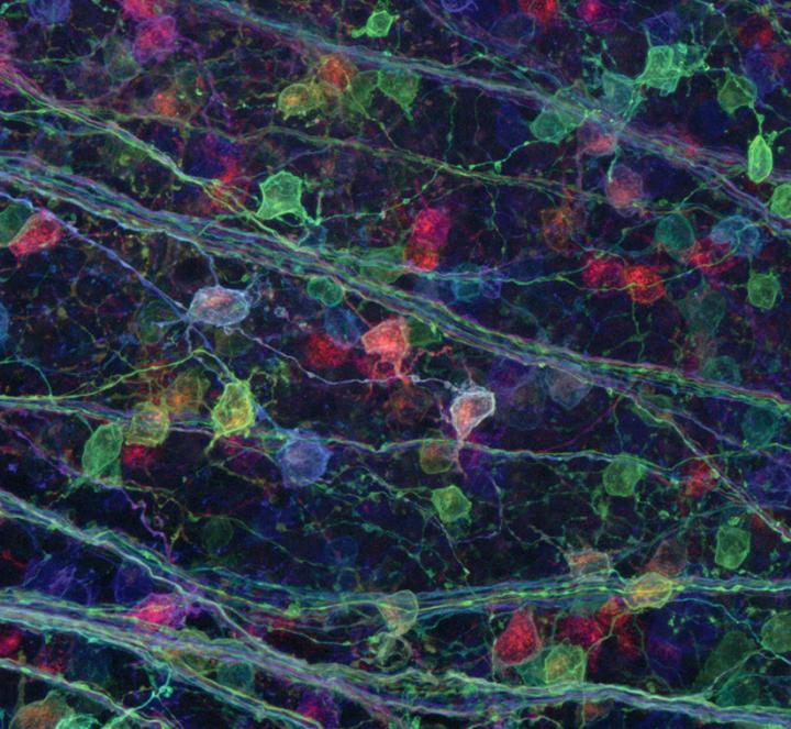
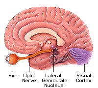
'Brainbow' reveals a surprising visual connection Neuroscientists know some connections in the brain are pruned during infant growth and neural development. According to the textbooks, function drives new neural growth. But scientists are discovering the textbooks might be wrong. "Retinal neurons associated with vision generate connections in the brain, and as the brain develops it strengthens and maintains some of those connections more than others, while unused connections are eliminated," says Michael Fox, Associate Professor at the Virginia Tech Carilion Research Institute and leader of the study. However, "We found that activity-dependent pruning might not be as simple as we believed." Their results were published in Cell Reports.
But as the theory of several terminals blossoming from one retinal ganglion cell had not yet been proved, Fox and his researchers decided to follow these terminals back to their roots. Using a technique dubbed "brainbow," the scientists tagged the terminals with fluorescent proteins of different colors, expecting one color to dominate. Instead, several colors appeared, intertwined and distinct from each other.
The results showed individual terminals come from more than one retinal ganglion in a mature mouse brain. This is in direct contradiction to previous research indicating neural development weeds out most connections between retinal ganglion and target cells in the brain. Fox: "Is this a discrepancy - a technical issue with the different types of approaches applied in all of these disparate studies? Possibly, but more likely it represents that retinal ganglion cells are more complex than previously thought." Along with the brainbow technique, Fox's team also imaged the connections with electron microscopy. Sarah Hammer, currently a sophomore at Virginia Tech, traced individual retinal terminals through hundreds of serial images. The data confirmed the "brainbow" analysis results - retinal axons from numerous retinal ganglion cells remained present on adult brain cells. "These results are not what we expected, and they will force us to reevaluate our understanding of the architecture and flow of visual information through neural pathways," Fox added. "The dichotomy of these results also sheds important light on the benefits of combining approaches to understand complicated problems in science."
Fox's team is continuing research to understand exactly how many retinal terminals converge and if the information each conveys is unique. Once they understand the intricacy of the brain's visual circuitry, the scientists might be able to develop therapies for when errors occur. Fox adds: "The lesson in this particular study is that no single technique gives us all the right answers. Science is never as simple as we'd like it to be." Abstract Highlights This is an open access article under the CC BY license (http://creativecommons.org/licenses/by/4.0/).
|
||||||||||||||||||||||||||||