|
|||||||||||||||
|

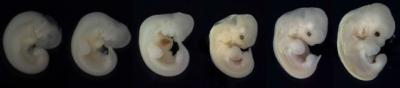
CLICK ON weeks 0 - 40 and follow along every 2 weeks of fetal development
|
||||||||||||||||||||||||||||
|
Fetal Timeline Maternal Timeline News News Archive Sep 14, 2015
|
Fats help organize spinal cord Healing spinal cord damage is incredibly difficult. Torn neurons must reconnect with precision, which we cannot make happen - as yet. But, scientists at the RIKEN Brain Science Institute in Japan have discovered that lipids are as needed for the process of guiding axons, as proteins are — a discovery which may improve our attempts.
Axons are long extensions of neurons, and act as roads along which neural information travels. Their paths are initiated in response to each of our senses, but eventually branch off and become more diffuse. During development, they are either attracted or repeled by molecules which force them along specic chanels and in specific directions along the spinal cord.
Before reaching our brains, sensory information is initiated through our skin and muscles to travel along our spinal cord. Axons carry these minute electrical impulses initially all in one surge, but soon separates them. Impulses responsible for our ability to sense pain — nociception — travel along the sides of the spinal cord, while those that tell us the position, direction, and equilibrium involved — proprioception — travel close to the middle of the spinal cord. Researchers were able to identify these pathways by experiments on chicken eggs. In a petri dish, they labeled chick spinal cord sections with molecular markers for LysoPtdGlc as well as for nociception and proprioception sensing neurons. They found LysoPtdGlc was located only near the midline of the spinal cord where proprioception or position-sensing axons are located.
To test this theory, they looked at how cultured pain-sensing nociception neurons responded to the LysoPtdGlc lipid. When introduced within sections of chick spinal cord, they observed that LysoPtdGlc repelled axons from nociception neurons. This was confirmed when they next blocked access to the LysoPtdGlc lipid with an experimental antibody and prevented any nociception or pain-sensing neurons from being repelled. The researchers then moved their experiments out of the petri dish and injected the antibody into the spinal cord of chick embryos. Their hypothesis that LysoPtdGlc was responsible for directing axon growth proved correct The axons of pain-sensing neurons were no longer repelled, and instead migrated into the region on the spinal cord reserved for position-sensitive neurons.
Having determinined that LysoPtdGlc's ability to repel pain-sensing nociception axons is controlled through a particular protein receptor on axons, the team tested over 100 receptors and found one — GPR55 — that responded well to LysoPtdGlc. This protein is also expressed in the spinal cord, and when the team labeled axons in mice with GPR55 turned off ( or supressed), they found pain-sensing axons mistakenly enter the upper-medial or middle portion of the spinal cord.
Abstract Reference: Guy, AT et al. Glycerophospholipid regulation of modality-specific sensory axon guidance in spinal cord. Science, doi:
|
||||||||||||||||||||||||||||