Welcome to The Visible Embryo
  o
 
The Visible Embryo Home
   
Google  
Home--- -History-----Bibliography-----Pregnancy Timeline-----Prescription Drugs in Pregnancy---- Pregnancy Calculator----Female Reproductive System----News----Contact
 
Welcome to The Visible Embryo, a comprehensive educational resource on human development from conception to birth.

The Visible Embryo provides visual references for changes in fetal development throughout pregnancy and can be navigated via fetal development or maternal changes.

The National Institutes of Child Health and Human Development awarded Phase I and Phase II Small Business Innovative Research Grants to develop The Visible Embryo. Initally designed to evaluate the internet as a teaching tool for first year medical students, The Visible Embryo is linked to over 600 educational institutions and is viewed by more than one million visitors each month.

Today, The Visible Embryo is linked to over 600 educational institutions and is viewed by more than 1 million visitors each month. The field of early embryology has grown to include the identification of the stem cell as not only critical to organogenesis in the embryo, but equally critical to organ function and repair in the adult human. The identification and understanding of genetic malfunction, inflammatory responses, and the progression in chronic disease, begins with a grounding in primary cellular and systemic functions manifested in the study of the early embryo.

WHO International Clinical Trials Registry Platform


The World Health Organization (WHO) has created a new Web site to help researchers, doctors and patients obtain reliable information on high-quality clinical trials. Now you can go to one website and search all registers to identify clinical trial research underway around the world!


Home

History

Bibliography

Pregnancy Timeline

Prescription Drug Effects on Pregnancy

Pregnancy Calculator

Female Reproductive System

News

Disclaimer: The Visible Embryo web site is provided for your general information only. The information contained on this site should not be treated as a substitute for medical, legal or other professional advice. Neither is The Visible Embryo responsible or liable for the contents of any websites of third parties which are listed on this site.


Content protected under a Creative Commons License.
No dirivative works may be made or used for commercial purposes.


Pregnancy Timeline by SemestersDevelopmental TimelineFertilizationFirst TrimesterSecond TrimesterThird TrimesterFirst Thin Layer of Skin AppearsEnd of Embryonic PeriodEnd of Embryonic PeriodFemale Reproductive SystemBeginning Cerebral HemispheresA Four Chambered HeartFirst Detectable Brain WavesThe Appearance of SomitesBasic Brain Structure in PlaceHeartbeat can be detectedHeartbeat can be detectedFinger and toe prints appearFinger and toe prints appearFetal sexual organs visibleBrown fat surrounds lymphatic systemBone marrow starts making blood cellsBone marrow starts making blood cellsInner Ear Bones HardenSensory brain waves begin to activateSensory brain waves begin to activateFetal liver is producing blood cellsBrain convolutions beginBrain convolutions beginImmune system beginningWhite fat begins to be madeHead may position into pelvisWhite fat begins to be madePeriod of rapid brain growthFull TermHead may position into pelvisImmune system beginningLungs begin to produce surfactant
CLICK ON weeks 0 - 40 and follow along every 2 weeks of fetal development


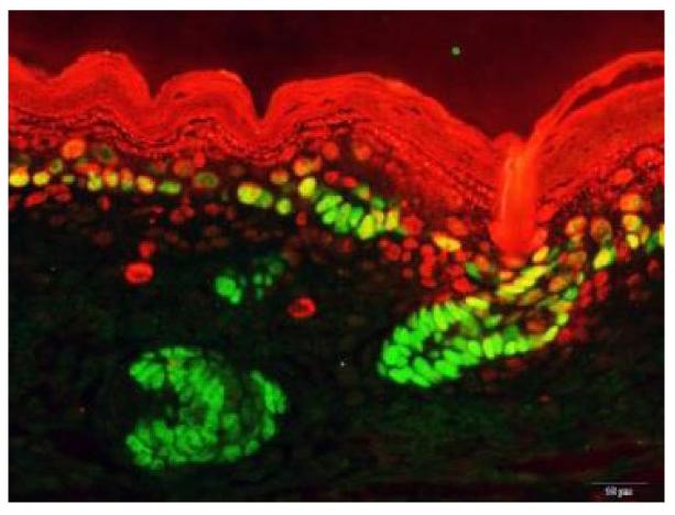
Two genes play major role in growth and cancers

Disruptions in the splicing of proteins causes facial, skin, and organ defects in young mice.


Knocking out one or both of these two crucial regulatory genes caused cleft lip, skin barrier defects, and a host of other problems in developing mice, according to research from the Perelman School of Medicine at the University of Pennsylvania. This outcome hints that abnormalities in molecular pathways underlie many birth defects.


Two regulatory genes, Esrp1 and Esrp2, are active in normal mammal development. They govern how RNAs, produced from genes, join to make the final version of protein in a process called alternative splicing.


Published this week in eLife, the work explains how two genes, Esrp1 and Esrp2, not only play a role in development - but also in cancers.

"Clearly there are many important roles for these genes in facial, skin and, organ development, and we're only just starting to catalogue them,"
said Russ P. Carstens, MD, an Associate Professor of Medicine (Renal-Electrolyte and Hypertension Division) and Genetics at the University of Pennsylvania.

Carstens and his colleagues in 2009 first identified Esrp1 and 2 as molecular switches for alternative splicing — a process in which RNA is transcribed from a gene's DNA. The DNA is cut up and put back together in different arrangements or "alternative splicings."  Most mammalian genes use it to enable one gene to produce many protein variations — called isoforms.

Which isoform is produced depends on the cell type and other physiologic circumstances. Esrp1 and Esrp2 are specific to epithelial cells which form skin, the inner lining of our gut and lungs, and some other tissues in the body. Carstens and his team since 2009 have shown that Esrp1 and 2 proteins can splice hundreds of different isoforms from epithelial-specific genes.

In 2013, they discovered that suppressing Esrp activity enables epithelial cells to detach and develop into new tissue — a process called epithelial-mesenchymal transition (EMT). Obviously EMT is crucial for creating new tissue during embry development and later for healing wounds.

But if abnormally switched on, EMT can enable the spread of tumor cells.

Early studies of the two proteins were made in laboratory-cultured cells. In this eLife study, Carstens and his colleagues have engineered mice without Esrp1 or Esrp2 or both. They found that mice born alive but without Esrp1, had cleft palate and cleft lip. Their deformities prevented feeding and all pups died within hours.


"This finding really shows that epithelial-specific splicing is important for the normal development of the face and palate — cleft lip and palate are among the most common congenital conditions."

Russ P. Carstens MD, Associate Professor of Medicine, Renal-Electrolyte and Hypertension Division, and Genetics.


Mice lacking only Esrp2 — which likely evolved as a backup copy to cover some functions of Esrp1 — appeared normal if Esrp1 was still present as a gene.

Mice lacking both genes had more abnormalities. Their embryos had more profound defects than in the mice missing Esrp1, including craniofacial and forelimb defects, and the complete absence of lungs and salivary glands — two organs largely from epithelial cells.

Collaborating with Yi Xing from UCLA, the team catalogued and analyzed how gene patterns in skin cells and how they differed among mice with various Esrp gene knockout combinations, finding hundreds of significant changes.

Their analysis suggested that the lack of both Esrp1 and Esrp2 leads to serious defects in skin development.

Because mice lacking both genes would not be born alive, the scientists made "conditional knockout mice," in which Esrp1 and Esrp2 activity was normal early in fetal development, but then was turned off in skin epithelial cells.

These mice perished within a day of being born. "They basically became dehydrated and died because their skin couldn't keep water and fluid in," said Carstens. This flaw is called an epithelial barrier defect, found in human disorders and includes the skin, gut, and lungs. Carstens hopes that more study of Esrp1 and Esrp2, particularly the proteins isoforms they control, will uncover clues to how these defects occur and how they might be corrected.

Carsten also hopes Esrp1-knockout mice will prove to be a valuable new model animal for studying cleft lip, adding: "There have been many knockout mouse models of cleft palate, but very few of cleft lip, which is actually the more common defect in humans."

The team is now working on other gene knockout studies of Esrp1 and Esrp2's as they affect face, lungs, gut, kidney, and other organs.

Abstract
Tissue- and cell-type-specific regulators of alternative splicing (AS) are essential components of posttranscriptional gene regulation, necessary for normal cellular function, patterning, and development. Mice with ablation of Epithelial splicing regulatory protein (Esrp1) develop cleft lip and palate. Loss of both Esrp1 and its paralog Esrp2 results in widespread developmental defects with broad implications to human disease. Deletion of the Esrps in the epidermis revealed their requirement for establishing a proper skin barrier, a primary function of epithelial cells comprising the epidermis. We profiled the global Esrp-mediated splicing regulatory program in epidermis, which revealed large-scale programs of epithelial cell-type-specific splicing required for epithelial cell functions. These mice represent a valuable model for evaluating the essential role for AS in development and function of epithelial cells, which play essential roles in tissue homeostasis in numerous organs, and provide a genetic tool to evaluate important functional properties of epithelial-specific splice variants in vivo.

DOI: http://dx.doi.org/10.7554/eLife.08954.001

See more at: http://elifesciences.org/content/4/e08954.full#sthash.e8r8JHqz.dpuf

The lead authors of this work were postdoctoral fellows Thomas W. Bebee, from Penn, who generated and characterized the knockout mice, who along with bioinformatic analysis by Juw Won Park, from UCLA, profiled the global molecular changes in the knockout mice. Other coauthors are Katherine Sheridan, Claude Warzecha, Benjamin Cieply, and Alex Rohacek, all from Penn.

Funding was provided by the National Institutes of Health (R01 GM088809, R56 AR066741, R56 DE024749, P30 AR057217, P30 AR050950, P30 DK050306, T32DK700638, F32DK098917, R01 GM088342, R01 GM105431), and a research fellowship to Yi Xing from the Alfred P. Sloan Foundation.

Penn Medicine is one of the world's leading academic medical centers, dedicated to the related missions of medical education, biomedical research, and excellence in patient care. Penn Medicine consists of the Raymond and Ruth Perelman School of Medicine at the University of Pennsylvania (founded in 1765 as the nation's first medical school) and the University of Pennsylvania Health System, which together form a $4.9 billion enterprise.

The Perelman School of Medicine has been ranked among the top five medical schools in the United States for the past 17 years, according to U.S. News & World Report's survey of research-oriented medical schools. The School is consistently among the nation's top recipients of funding from the National Institutes of Health, with $409 million awarded in the 2014 fiscal year.

The University of Pennsylvania Health System's patient care facilities include: The Hospital of the University of Pennsylvania and Penn Presbyterian Medical Center -- which are recognized as one of the nation's top "Honor Roll" hospitals by U.S. News & World Report -- Chester County Hospital; Lancaster General Health; Penn Wissahickon Hospice; and Pennsylvania Hospital -- the nation's first hospital, founded in 1751. Additional affiliated inpatient care facilities and services throughout the Philadelphia region include Chestnut Hill Hospital and Good Shepherd Penn Partners, a partnership between Good Shepherd Rehabilitation Network and Penn Medicine.


Return to top of page

Sep 21, 2015   Fetal Timeline   Maternal Timeline     News     News Archive   


Immunofluorescence of skin reflected in basal cells.
Image Credit: Russ Carstens, M.D., Perelman School
of Medicine, University of Pennsylvania; eLifeImage K


Phospholid by Wikipedia