|
|
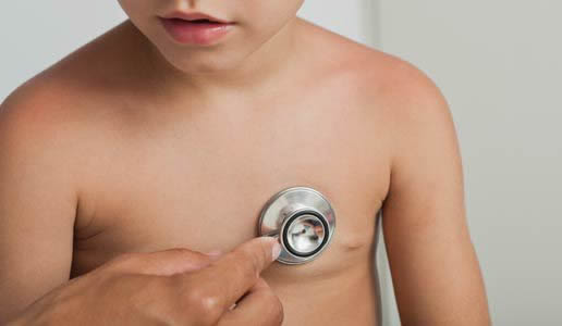
Mom's protein deficiency affects boys more
A study has uncovered the genetic processes that link insufficient protein during pregnancy with problems in muscle development in boy babies.
The findings also reveal the metabolic pathway through which genetic changes are transferred to the fetus, potentially triggering development of chronic health problems such as cardiovascular disease, obesity and Type 2 diabetes in the latter adult, according to research conducted at the University of Illinois.
Huan Wang PhD, the principal investigator on the study, detected biomarkers of protein insufficiency in mothers during their early stages of pregnancy. Her observations could lead clinicians to treat such an insufficiency through diet or some other strategy, and possibly avert serious health problems in her developing fetus.
Her work is published in the British Journal of Nutrition. Although Wang used rats as her model animal in this particular study, prior research has shown that rat physiology is quite similar to human physiology and the implications from the study apply to humans.
During pregnancy, women require an additional 25 grams of protein per day, at least. Wang found that inadequate protein levels during pregnancy activates an Amino Acid Response (AAR) molecular pathway, which triggers cell destruction - or autophagy - as well as atrophy, or wasting of the mother's skeletal muscles.
Autophagy is a survival mechanism. Cells under stress degrade unneeded or dysfunctional parts of the cell in order to maintain a cell's stability and consistency — and of course the tissue made up of those cells benefits. However, autophagy out of balance can negatively affect the whole body as damaged muscle fibers may fail to quickly contract and expand when needed.
These genetic changes brought on by the amino acid response (AAR) appear to migrate through the placenta and become "memorized" in the skeletal muscles of the fetus — causing low weight at birth, as well as stunted growth in male babies.
"This is the link we've been seeking for years, it shows transduction from the mom through the placenta to the child. However, cell autophagy is activated in the skeletal muscles of the male child only. Apparently, female offspring have more resistance to low-protein exposure and cell autophagy during gestation."
Huan Wang PhD, who conducted her research while completing a doctorate in food science and human nutrition at the University of Illinois. She is currently a postdoctoral researcher in human genetics at the University of California at Los Angeles.
For the study, pregnant rats were fed food containing 8 to 9 percent protein. However, those in a control group ate twice that much - 18 to 20 percent protein. After giving birth, all of the rats ate 18 to 20 percent protein in their diet when nursing. Also, all of the pups after being weaned from their mothers also ate 18 to 20 percent protein in their diet.
All the rats' weights and food intake were recorded every other day, and revealed that mother rats on the low-protein diet gained significantly less weight during pregnancy — and their pups were smaller at birth as well. The low-protein diet also changed the levels of key amino acids in the mothers' blood plasma.
At the end of pregnancy, mothers in the low-protein group had lower levels of threonine and histidine, and higher levels of alanine, lysine and serine, which suggest potential disturbances in their protein metabolism. Examining the moms' skeletal muscle fibers after delivery also revealed muscle atrophy, including smaller muscle fiber size, greater variation in muscle fiber diameter, as well as split fibers. Again, female pups, were not affected.
Mothers on the low-protein diet showed higher expression of the gene ATF4, a key regulatory protein within the Amino Acid Response (AAR) pathway. ATF4 was recently found to be critical in muscle dystrophy caused by fasting, and is associated with cell autophagy (cell death).
The turning on of autophagy genes and the binding of these genes to ATF4 confirms a molecular link between them and the amino acid response (AAR) signal. Follow-up data indicating that muscle deterioration remained active in adult males deprived of protein during their moms' pregnancy — supports the concept that the AAR signal transfers through the placenta to the fetus.
Altogether, this research underscores how important it is pregnant women eat enough protein to assist fetal muscle formation that will ultimately affect their male children as adults.
Abstract
The aim of the present study was to investigate the mechanistic basis of protein deficiency during pregnancy in mother that is transduced to offspring. To this end, timed-pregnant Sprague–Dawley rats were fed either a control (20 % of energy from protein) or low-protein (LP, 8 % of energy from protein) diet during gestation. Tissues were collected after delivery from rat dams, and skeletal muscle was collected at postnatal day 38 from the offspring. Quantitative RT-PCR and Western blot analyses were performed to determine mRNA and protein levels. Histological analysis was performed to evaluate myofibre size. LP dams gained significantly less weight during pregnancy, developed muscle atrophy, and had significantly lower circulating threonine and histidine levels than control dams. The mRNA expression of the well-known amino acid response (AAR) pathway-related target genes was increased only in the skeletal muscle of LP dams, as well as the protein expression levels of activating transcription factor 4 (ATF4) and phosphorylated eukaryotic translation initiation factor 2α (p-eIF2α). The mRNA expression of autophagy-related genes was significantly increased in the skeletal muscle of LP dams. Moreover, the mRNA expression of genes involved in both AAR and autophagy pathways remained elevated and was memorised in the muscle of LP offspring that consumed a post-weaning control diet. Additionally, the LP diet increased an autophagy marker, microtubule-associated proteins 1A/1B light chain 3B (LC3B) protein expression in the skeletal muscle of rat dams, consistent with the initiation of autophagy. The LP diet further increased ATF4 binding at the predicted regions of AAR and autophagy pathway-related genes. Increased binding of ATF4 unveils the crucial role of ATF4 in the activation of autophagy in response to protein restriction. Our data suggest that molecular changes in maternal muscle are memorised in the offspring long after gestational protein restriction, reinforcing the role of maternal signalling in programming offspring health.
Wang's co-authors on the paper were U. of I. faculty members Yuan-Xiang Pan and Donald K. Layman, both in food science; and Stéphane Lezmi, nutritional sciences. Huan Wang is currently a postdoctoral researcher in human genetics at the University of California at Los Angeles.
Illinois alumni Xiuwen Chen, Gabriel J. Wilson and Dan Zhou also were co-authors on the paper.
Return to top of page
|
|
|
Sep 29, 2015 Fetal Timeline Maternal Timeline News News Archive
This research suggest pregnant women increase protein in their diet
to avoid muscle decrease in their babies, particularly in male children.
.Image Credit: public domain stock photo
|
|
| |
|


