|
|||||||||||||||
|

CLICK ON weeks 0 - 40 and follow along every 2 weeks of fetal development
|
||||||||||||||||||||||||||||
|
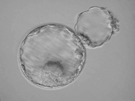
Splitting embryo to force twins for IVF - not advised Human twin embryos created in the laboratory, by splitting single embryos into two, is a common method known as blastomere biopsy. But a new study led by King's College London, suggests this split may be interferring with a 'developmental clock' critical to early human development. Therefore, unsuitable for either research or IVF. In the UK, the Human Fertilisation and Embryology Authority Code of Practice makes it clear that clinics should not be producing embryos for In Vitro Fertilization (IVF) treatment by embryo splitting. They advise such genetically identical embryos should be used only for research purposes. The latest study, led by PhD student Laila Noli from King's College London and published in the journal Human Reproduction, set out to determine whether the quality of human embryos generated by twinning ' by in-vitro' (in the laboratory) was comparable to the quality of embryos created by fertilization of eggs by sperm done for IVF. Using time lapse cameras for monitoring, researchers compared the development of 176 twin embryos created by manually splitting 88 human embryos at two different stages, early (2–5 blastomeres) or late (6–10 blastomeres) cleavage. Under examination, researchers found delays in the development of embryos generated by manipulated twinning, compared to fertilized singleton embryos created through IVF.
Abstract SUMMARY ANSWER Our data suggest that the human twin embryos created in vitro are unsuitable not only for clinical use but also for research purposes. WHAT IS KNOWN ALREADY Pregnancy from in vitro generated monozygotic twins by embryo splitting or twinning leads to live birth of healthy animals. Similar strategies, however, have been less successful in primates. Recent reports suggest that the splitting of human embryos might result in viable, morphologically adequate blastocysts, although the qualitative analyses of the embryos created in such a way have been very limited. STUDY DESIGN, SIZE, DURATION This study was a comparative analysis of embryos generated by twinning in vitro and the embryos created by in vitro fertilization. PARTICIPANTS/MATERIALS, SETTING, METHODS We analysed morphokinetics and developmental competence of 176 twin embryos created by splitting of 88 human embryos from either early (2–5 blastomeres, n = 43) or late (6–10 blastomeres, n = 45) cleavage stages. We compared the data with morphometrics of embryos created by in vitro fertilization and resulting in pregnancy and live birth upon single blastocyst transfer (n = 42). MAIN RESULTS AND THE ROLE OF CHANCE The morphokinetic data suggested that the human preimplantation development is subjected to a strict temporal control. Due to a ‘developmental clock’, the size of twin embryos was proportionate to the number of cells used for their creation. Furthermore, the first fate decision was somewhat delayed; the inner cell mass (ICM) became distinguishable later in the twin than in the normal blastocysts obtained through fertilization. If an ICM was present at all, it was small and of poor quality. The majority of the cells in the twin embryos expressed ICM and trophectoderm markers simultaneously. LIMITATIONS, REASONS FOR CAUTION We created monozygotic twins by blastomere separation from cleavage stage embryos. Embryo twinning by blastocyst bisection may circumvent limitations set by the developmental clock. WIDER IMPLICATIONS OF THE FINDINGS Taken together, our data suggest that the human twin embryos created in vitro are unsuitable not only for clinical use but also for research purposes. STUDY FUNDING/COMPETING INTEREST(S) This project was supported by the Saudi Arabian Government studentship to L.N. and incentive funds to Y.K. and D.I. The authors have no potential conflict of interest. |
Oct 27, 2015 Fetal Timeline Maternal Timeline News News Archive
|
||||||||||||||||||||||||||||


