Welcome to The Visible Embryo
  o
 
The Visible Embryo Home
   
Google  
Home--- -History-----Bibliography-----Pregnancy Timeline-----Prescription Drugs in Pregnancy---- Pregnancy Calculator----Female Reproductive System----News----Contact
 
Welcome to The Visible Embryo, a comprehensive educational resource on human development from conception to birth.

The Visible Embryo provides visual references for changes in fetal development throughout pregnancy and can be navigated via fetal development or maternal changes.

The National Institutes of Child Health and Human Development awarded Phase I and Phase II Small Business Innovative Research Grants to develop The Visible Embryo. Initally designed to evaluate the internet as a teaching tool for first year medical students, The Visible Embryo is linked to over 600 educational institutions and is viewed by more than one million visitors each month.

Today, The Visible Embryo is linked to over 600 educational institutions and is viewed by more than 1 million visitors each month. The field of early embryology has grown to include the identification of the stem cell as not only critical to organogenesis in the embryo, but equally critical to organ function and repair in the adult human. The identification and understanding of genetic malfunction, inflammatory responses, and the progression in chronic disease, begins with a grounding in primary cellular and systemic functions manifested in the study of the early embryo.

WHO International Clinical Trials Registry Platform


The World Health Organization (WHO) has created a new Web site to help researchers, doctors and patients obtain reliable information on high-quality clinical trials. Now you can go to one website and search all registers to identify clinical trial research underway around the world!


Home

History

Bibliography

Pregnancy Timeline

Prescription Drug Effects on Pregnancy

Pregnancy Calculator

Female Reproductive System

News

Disclaimer: The Visible Embryo web site is provided for your general information only. The information contained on this site should not be treated as a substitute for medical, legal or other professional advice. Neither is The Visible Embryo responsible or liable for the contents of any websites of third parties which are listed on this site.


Content protected under a Creative Commons License.
No dirivative works may be made or used for commercial purposes.


Pregnancy Timeline by SemestersDevelopmental TimelineFertilizationFirst TrimesterSecond TrimesterThird TrimesterFirst Thin Layer of Skin AppearsEnd of Embryonic PeriodEnd of Embryonic PeriodFemale Reproductive SystemBeginning Cerebral HemispheresA Four Chambered HeartFirst Detectable Brain WavesThe Appearance of SomitesBasic Brain Structure in PlaceHeartbeat can be detectedHeartbeat can be detectedFinger and toe prints appearFinger and toe prints appearFetal sexual organs visibleBrown fat surrounds lymphatic systemBone marrow starts making blood cellsBone marrow starts making blood cellsInner Ear Bones HardenSensory brain waves begin to activateSensory brain waves begin to activateFetal liver is producing blood cellsBrain convolutions beginBrain convolutions beginImmune system beginningWhite fat begins to be madeHead may position into pelvisWhite fat begins to be madePeriod of rapid brain growthFull TermHead may position into pelvisImmune system beginningLungs begin to produce surfactant
CLICK ON weeks 0 - 40 and follow along every 2 weeks of fetal development


Mom's preeclampsia increases risk to baby's heart

Pregnant women with preeclampsia have a higher risk of delivering an infant with a congenital heart defect.


An extensive study of 1.9 million mother and infant pairs at the University of Montreal Hospital Research Centre (CRCHUM) has shown significant association between preeclampsia in mothers and their newborns from early pregnancy.

The study is published in the prestigious Journal of the American Medical Association (JAMA),and is the basis for future research to detect and prevent these diseases.

Congenital heart defects are the most common anomalies in infants, affecting every 8 births in a 1,000. They are a major cause of infant illness and death, despite significant medical care advancements.

"We discovered that congenital heart defects were significantly more frequent for infants of women with preeclampsia during pregnancy," said Dr. Nathalie Auger, first author of the study, researcher at the CRCHUM, and professor at the University of Montreal's Department of Social and Preventive Medicine.

The origin of heart defects is sometimes hereditary, but risk factors were mostly unknown. Researchers had considered a possible link with preeclampsia, a pregnancy disorder characterized by high blood pressure and excess protein in the urine (proteinuria).


"Pregnant women with preeclampsia and infants born with heart defects have similarities in the biological formation of their blood vessels. This coincidence in the imbalance of angiogenic factors [growth factors that activate receptors on endothelial cells in blood vessels] prompted us to conduct this study."

Nathalie Auger MD, MSc, FRCPC, professor at the Department of Social and Preventive Medicine, University of Montreal Hospital Research Centre (CRCHUM), Montreal, Quebec, Canada, and first author of the study.


Researchers decided to examine hospital records of live births for the entire province of Quebec - making up of a quarter of Canada's population - between 1989 and 2012. All women who delivered an infant after 20 weeks of pregnancy during this period were included — and 1,942,072 neonates.

Overall, 17,296 neonates or 8.9 per 1,000, had heart defects. Prevalence was higher for infants of women with preeclampsia than without (16.7 vs. 8.6 per million). The most frequent heart defects were non-critical, such as septal defects, characterized by a hole in the wall separating the ventricles or atria of the heart and causing blood flow problems.


"The absolute risk of congenital heart defects is low. But there is a link between preeclampsia and heart defects in newborns. And the risk is higher when preeclampsia occurs before 34 weeks of pregnancy."

Nathalie Auger MD


Preeclampsia is detectable around the 20th week of pregnancy and can progress to full eclampsia endangering the life of the mother and child.

In fact, biomarkers that prevent the formation of blood vessels are present from the beginning of pregnancy in women who later develop preeclampsia.

The baby's heart is formed at the beginning of intrauterine life. Heart defects usually occur between eight and ten weeks of gestation. "There is a possible common factor in the embryo and mother that triggers both preeclampsia and heart defects very early in pregnancy," said the epidemiologist.

Currently, women diagnosed with preeclampsia are closely monitored to see if their baby is developing normally. Most infants with congenital heart defects can be saved. They are treated with medications, non-invasive procedures, or surgery.


"Further research is needed before changing medical recommendations.

"But knowing preeclampsia can increase the risk of congenital heart defects in an unborn child should help us better understand the pathophysiology of these diseases and ultimately improve their prevention and treatment."


Nathalie Auger MD


Abstract
Importance The risk of congenital heart defects in infants of women who had preeclampsia during pregnancy is poorly understood, despite shared angiogenic pathways in both conditions.

Objective To determine the prevalence of congenital heart defects in offspring of women with preeclampsia.

Design, Setting, and Participants Population-level analysis of live births before discharge, 1989-2012, was conducted for the entire province of Quebec, comprising a quarter of Canada’s population. All women who delivered an infant with or without heart defects in any Quebec hospital were included (N = 1?942?072 neonates).

Exposures Preeclampsia or eclampsia with onset before or after 34 weeks of gestation.

Main Outcomes and Measures Presence of any critical or noncritical congenital heart defect detected in infants at birth, comparing prevalence in those exposed and not exposed to preeclampsia.

Results The absolute prevalence of congenital heart defects was higher for infants of women with preeclampsia (16.7 per 1000 [1219/72?782]) than without it (8.6 per 1000 [16?077/1?869?290]; prevalence ratio [PR], 1.57; 95% CI, 1.48 to 1.67). Infants of preeclamptic women had no increased prevalence of critical heart defects (123.7 vs 75.6 per 100?000 [90/72?782 vs 1414/1?869?290]; PR, 1.25; 95% CI, 1.00 to 1.57; prevalence difference [PD], 23.6 per 100?000; 95% CI, −1.0 to 48.2) but did have an increased prevalence of noncritical heart defects (1538.8 vs 789.2 per 100?000 [1120/72?782 vs 14?752/1?869?290]; PR, 1.56; 95% CI, 1.47 to 1.67; PD, 521.1 per 100?000; 95% CI, 431.1 to 611.0) compared with infants of nonpreeclamptic women. Among specific defects, prevalence was greatest for septal defects. When stratified by variant of preeclampsia, infants of women with early onset (<34 weeks) preeclampsia had greater prevalence of critical heart defects (364.4 per 100?000 [20/5488]; PR, 2.78; 95% CI, 1.71-4.50; PD, 249.6 per 100?000; 95% CI, 89.7-409.6) and noncritical heart defects (7306.9 per 100?000 [401/5488]; PR, 5.55; 95% CI, 4.98-6.19; PD, 6089.2 per 100?000; 95% CI, 5350.0-6828.3), whereas infants of women with late onset (≥34 weeks) did not.

Conclusions and Relevance In this population-based study, preeclampsia was significantly associated with noncritical heart defects in offspring, and preeclampsia before 34 weeks was associated with critical heart defects. However, the absolute risk of congenital heart defects was low.

The study "Association Between Preeclampsia and Congenital Heart Defects," was published online in JAMA, The Journal of the American Medical Association, on October 20. The study was supported by the Canadian Institutes of Health Research (CIHR) (MOP-130452). Dr. Auger was supported by a career award from the Fonds de recherche du Québec - Santé (FRQS) (Grant 25128). The authors state that the funding organizations were not involved in the design and conduct of the study or in the publication of the results. For more information: http://jama.jamanetwork.com/article.aspx?articleid=2463255

Return to top of page

Oct 29, 2015   Fetal Timeline   Maternal Timeline   News   News Archive   


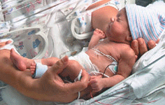
An extensive study of mothers and infant pairs has shown significant association
between preeclampsia in mothers and  heart disease in their newborns.
Image Credit: Public domain


Phospholid by Wikipedia