Welcome to The Visible Embryo
  o
 
The Visible Embryo Home
   
Google  
Home--- -History-----Bibliography-----Pregnancy Timeline-----Prescription Drugs in Pregnancy---- Pregnancy Calculator----Female Reproductive System----News----Contact
 
Welcome to The Visible Embryo, a comprehensive educational resource on human development from conception to birth.

The Visible Embryo provides visual references for changes in fetal development throughout pregnancy and can be navigated via fetal development or maternal changes.

The National Institutes of Child Health and Human Development awarded Phase I and Phase II Small Business Innovative Research Grants to develop The Visible Embryo. Initally designed to evaluate the internet as a teaching tool for first year medical students, The Visible Embryo is linked to over 600 educational institutions and is viewed by more than one million visitors each month.

Today, The Visible Embryo is linked to over 600 educational institutions and is viewed by more than 1 million visitors each month. The field of early embryology has grown to include the identification of the stem cell as not only critical to organogenesis in the embryo, but equally critical to organ function and repair in the adult human. The identification and understanding of genetic malfunction, inflammatory responses, and the progression in chronic disease, begins with a grounding in primary cellular and systemic functions manifested in the study of the early embryo.

WHO International Clinical Trials Registry Platform


The World Health Organization (WHO) has created a new Web site to help researchers, doctors and patients obtain reliable information on high-quality clinical trials. Now you can go to one website and search all registers to identify clinical trial research underway around the world!


Home

History

Bibliography

Pregnancy Timeline

Prescription Drug Effects on Pregnancy

Pregnancy Calculator

Female Reproductive System

News

Disclaimer: The Visible Embryo web site is provided for your general information only. The information contained on this site should not be treated as a substitute for medical, legal or other professional advice. Neither is The Visible Embryo responsible or liable for the contents of any websites of third parties which are listed on this site.


Content protected under a Creative Commons License.
No dirivative works may be made or used for commercial purposes.


Pregnancy Timeline by SemestersDevelopmental TimelineFertilizationFirst TrimesterSecond TrimesterThird TrimesterFirst Thin Layer of Skin AppearsEnd of Embryonic PeriodEnd of Embryonic PeriodFemale Reproductive SystemBeginning Cerebral HemispheresA Four Chambered HeartFirst Detectable Brain WavesThe Appearance of SomitesBasic Brain Structure in PlaceHeartbeat can be detectedHeartbeat can be detectedFinger and toe prints appearFinger and toe prints appearFetal sexual organs visibleBrown fat surrounds lymphatic systemBone marrow starts making blood cellsBone marrow starts making blood cellsInner Ear Bones HardenSensory brain waves begin to activateSensory brain waves begin to activateFetal liver is producing blood cellsBrain convolutions beginBrain convolutions beginImmune system beginningWhite fat begins to be madeHead may position into pelvisWhite fat begins to be madePeriod of rapid brain growthFull TermHead may position into pelvisImmune system beginningLungs begin to produce surfactant
CLICK ON weeks 0 - 40 and follow along every 2 weeks of fetal development


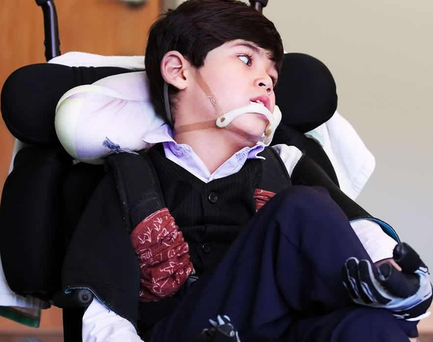
Batten disease may benefit from gene therapy

Batten disease causes problems with a cell's ability to breakdown specific molecules. In a study with dogs, scientists found a new way to deliver replacement genes effective at slowing the development of this rare and fatal childhood neurological disorder.


Early symptoms of Batten disease, a lysosomal storage disorder, may include vision loss, subtle personality and behavior changes, slow learning, clumsiness or stumbling. Eventually, the child becomes blind, bedridden and demented. Death usually comes within the first decade of life. There are no effective treatments.

Scientists have found a key to slowing down the disorder may be to inject viruses carrying the proper codes for needed gene products into the brain ventricles (see image below), the fluid-filled compartments in the center of the brain that serve as its plumbing system.

The study is published in Science Translational Medicine, and was partially funded by the National Institutes of Health.

"Our study opens up the possibility of a one-and-done treatment for this form of Batten disease," said Beverly Davidson, Ph.D., director of the Raymond G. Perelman Center for Cellular and Molecular Therapeutics at Children's Hospital of Philadelphia and the senior author of the study.

Working with scientists at the University of Missouri, Columbia, Davidson's team focused on the late infantile form of the disease that starts in children 2 to 4 years of age. It is most often caused by mutations in the gene for soluble lysosomal enzyme tripeptidyl peptidase 1 (TPP1) — an enzyme which degrades proteins.


Scientists found that if they treated dogs with a similar disorder, injecting a safe virus containing TPP1 gene code into cerebrospinal fluid that fills brain ventricles, the dogs lived about twice as long as untreated dogs.


Symptoms including problems with movement, pupil dilation and making decisions were delayed or in some cases did not occur. The treatment, however, did not fully improve a dogs' vision suggesting that delivery to the eye itself may be necessary as well. When the brains were inspected, the treatment had reduced damage normally caused by the disease. In comparison with untreated dogs, treated dogs had less reactive glial cells or stored lipofuscins — fatty deposits that are hallmarks of Batten disease and similar disorders.

"Dr. Davidson and her team undertook a highly innovative approach for Batten disease gene therapy," said Jill Morris, Ph.D., program director at NIH's National Institute of Neurological Disorders and Stroke. "These results open up a promising path toward developing long-lasting treatments for Batten disease and similar lysosomal storage disorders."


Further inspections confirmed their hypothesis.

Parenchymal cells lining ventricles and surface of the brain absorbed injected genes from cere-brospinal fluid and made more TPP1 protein.

The cells then secreted the TPP1 protein, spreading it throughout the brain.
  Brain Ventricles
Image credit: aps Physiological Reviews

Initial experiments showed that it was important to treat the dogs with an immunosuppressant — mycophenolate mofetil — before injecting the TPP1 gene. The immunosuppressant prevented the body from producing antibodies to clear TPP1 from the cerebrospinal fluid.

"We saw profound effects from the gene therapy that, summed up, improved the dogs' health. We certainly hope that this approach will provide children suffering from this disorder similar benefits," said Dr. Davidson.

Abstract
Repeated enzyme replacement therapy via infusion of the enzyme into blood vessels is an established treatment modality for many lysosomal storage diseases but not for those with significant brain involvement. In new work, Katz and colleagues test gene delivery of the missing enzyme to cells that line the fluid-filled spaces of the brain and measure secretion of the enzyme into the cerebrospinal fluid. Using a dog model of a lysosomal storage disease, they show that this strategy delayed disease onset, extended life span, and protected dogs from early cognitive decline, suggesting that this approach could improve the lives of children suffering from the same or similar diseases.

References: Katz, Tecedor, Chen et al. "AAV gene transfer delays disease onset in a TPP1-deficient canine model of the late infantile form of Batten disease," Science Translational Medicine, November 11, 2015. DOI: 10.10.1126/scitranslmed.aac6191

For more information, visit: http://www.ninds.nih.gov
http://www.ninds.nih.gov/disorders/batten/batten.htm
nei.nih.gov/

This work was supported by grants from NIH (NS068099, NS077516, EY023968), the Batten Disease Support and Research Association, Jasper Against Batten Fund at Partnership for Cures, Blake's Purpose Foundation, Roy J. Carver Trust, Children's Hospital of Philadelphia Research Institute.

The NINDS is the nation's leading funder of research on the brain and nervous system. The mission of NINDS is to seek fundamental knowledge about the brain and nervous system and to use that knowledge to reduce the burden of neurological disease.

NEI leads the federal government's research on the visual system and eye diseases. NEI supports basic and clinical science programs that result in the development of sight-saving treatments. For more information, visit http://www.nei.nih.gov.

About the National Institutes of Health (NIH): NIH, the nation's medical research agency, includes 27 Institutes and Centers and is a component of the U.S. Department of Health and Human Services. NIH is the primary federal agency conducting and supporting basic, clinical, and translational medical research, and is investigating the causes, treatments, and cures for both common and rare diseases. For more information about NIH and its programs, visit http://www.nih.gov.

Return to top of page

Nov 13, 2015   Fetal Timeline   Maternal Timeline   News   News Archive   


Batten disease is a rare, fatal autosomal-recessive neurodegenerative disorder named for
the British pediatrician who identified the condition in 1903. It is the most common form
of a group of disorders called neuronal ceroid lipofuscinoses (NCLs).

Image Credit: The Medical Bag


Phospholid by Wikipedia