Welcome to The Visible Embryo
  o
 
The Visible Embryo Home
   
Google  
Home--- -History-----Bibliography-----Pregnancy Timeline-----Prescription Drugs in Pregnancy---- Pregnancy Calculator----Female Reproductive System----News----Contact
 
Welcome to The Visible Embryo, a comprehensive educational resource on human development from conception to birth.

The Visible Embryo provides visual references for changes in fetal development throughout pregnancy and can be navigated via fetal development or maternal changes.

The National Institutes of Child Health and Human Development awarded Phase I and Phase II Small Business Innovative Research Grants to develop The Visible Embryo. Initally designed to evaluate the internet as a teaching tool for first year medical students, The Visible Embryo is linked to over 600 educational institutions and is viewed by more than one million visitors each month.

Today, The Visible Embryo is linked to over 600 educational institutions and is viewed by more than 1 million visitors each month. The field of early embryology has grown to include the identification of the stem cell as not only critical to organogenesis in the embryo, but equally critical to organ function and repair in the adult human. The identification and understanding of genetic malfunction, inflammatory responses, and the progression in chronic disease, begins with a grounding in primary cellular and systemic functions manifested in the study of the early embryo.

WHO International Clinical Trials Registry Platform


The World Health Organization (WHO) has created a new Web site to help researchers, doctors and patients obtain reliable information on high-quality clinical trials. Now you can go to one website and search all registers to identify clinical trial research underway around the world!


Home

History

Bibliography

Pregnancy Timeline

Prescription Drug Effects on Pregnancy

Pregnancy Calculator

Female Reproductive System

News

Disclaimer: The Visible Embryo web site is provided for your general information only. The information contained on this site should not be treated as a substitute for medical, legal or other professional advice. Neither is The Visible Embryo responsible or liable for the contents of any websites of third parties which are listed on this site.


Content protected under a Creative Commons License.
No dirivative works may be made or used for commercial purposes.


Pregnancy Timeline by SemestersDevelopmental TimelineFertilizationFirst TrimesterSecond TrimesterThird TrimesterFirst Thin Layer of Skin AppearsEnd of Embryonic PeriodEnd of Embryonic PeriodFemale Reproductive SystemBeginning Cerebral HemispheresA Four Chambered HeartFirst Detectable Brain WavesThe Appearance of SomitesBasic Brain Structure in PlaceHeartbeat can be detectedHeartbeat can be detectedFinger and toe prints appearFinger and toe prints appearFetal sexual organs visibleBrown fat surrounds lymphatic systemBone marrow starts making blood cellsBone marrow starts making blood cellsInner Ear Bones HardenSensory brain waves begin to activateSensory brain waves begin to activateFetal liver is producing blood cellsBrain convolutions beginBrain convolutions beginImmune system beginningWhite fat begins to be madeHead may position into pelvisWhite fat begins to be madePeriod of rapid brain growthFull TermHead may position into pelvisImmune system beginningLungs begin to produce surfactant
CLICK ON weeks 0 - 40 and follow along every 2 weeks of fetal development


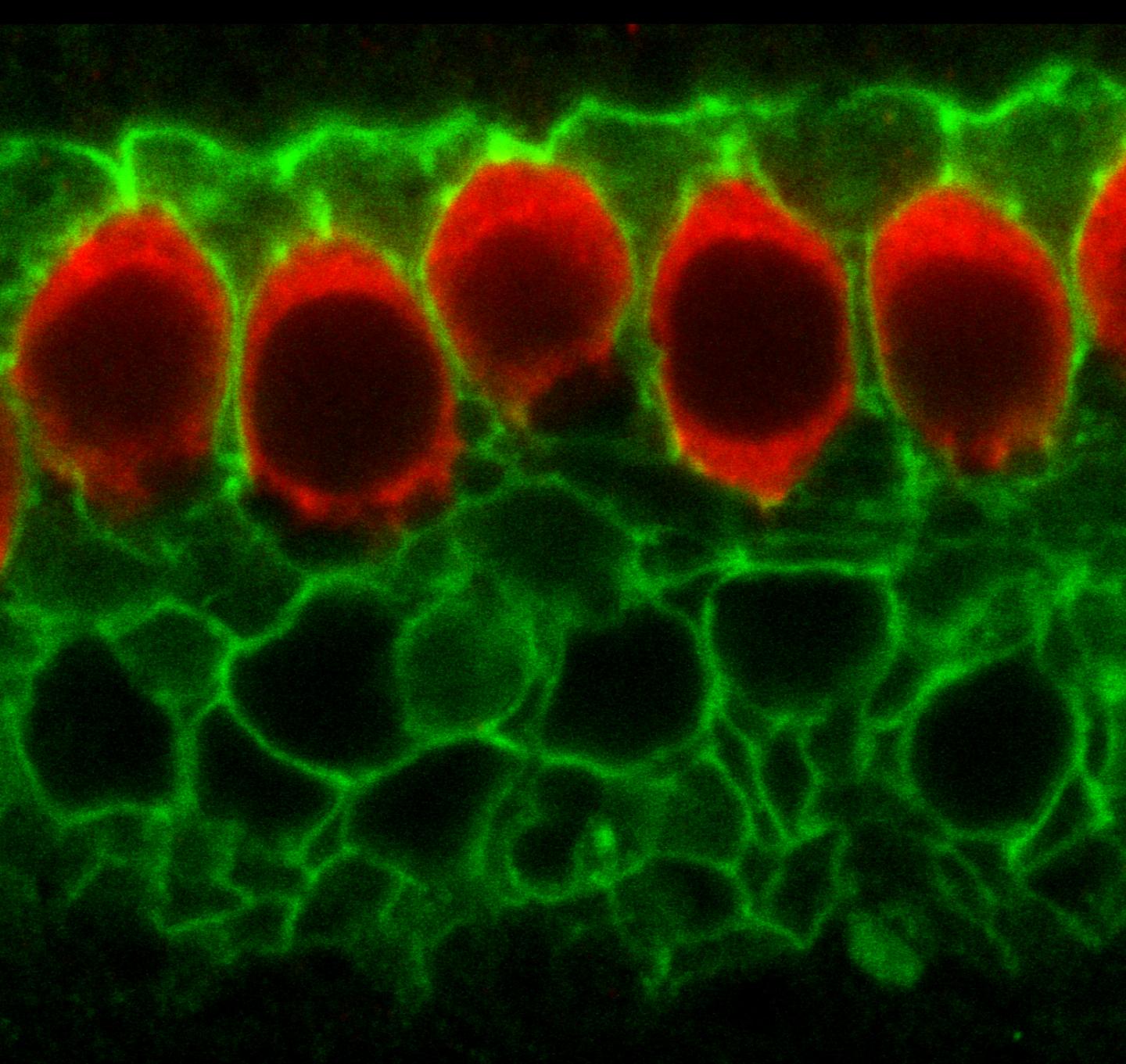
How cells in the developing ear 'practice' hearing

Before the fluid of the middle ear drains and sound waves penetrate those cells for the first time, inner ear cells of newborn rodents practice for their big debut.


Researchers at Johns Hopkins report they have figured out the molecular chain of events that enables cells to make "sounds" on their own, essentially "practicing" how to process sound in the world around them. Describing their experiments in the December edition of the journal Cell, scientists show how hair cells in the inner ear can be activated in the absence of sound.

"The multistep process we uncovered reminds me of a Rube Goldberg invention. Cells in the inner ear exploit a system used for fluid secretion in other organs to simulate the effect of sound before hearing begins, preparing them for the real deal," says Dwight Bergles PhD, professor of neuroscience at the Johns Hopkins University School of Medicine, in a reference to Rube Goldberg's construction of complicated gadgets to perform seemingly simple tasks.


Normal hearing in most mammals is a multistep process. It begins with sound waves hitting the ear drum, which transfers that energy into the air-filled middle ear and its three tiny bones.

When fluid in the inner ear vibrates at an electrical frequency, "antennae" on just the right "hair cells" bend causing them to release a chemical message to nearby nerves to fire.

That nerve signal travels to the brain, where it is interpreted as a particular sound.


Scientists, Bergles says, already knew that hair cells and nearby "supporting" cells in the developing inner ear show synchronous bursts of activity triggered by release of the chemical ATP — also used as a potent communication signal. This activity is then conveyed to the brain in the same way that sound-evoked information is, leading to burst firing of neurons in different auditory centers of the brain.

What was unknown was how ATP activates hair cells.

To find out, Bergles and his team honed in on biochemical elements of the system and found that chloride ion channels in supporting cells appeared crucial. They knew that ATP triggers a rise in calcium levels inside supporting cells, so they guessed that calcium was the signal for a calcium-activated chloride channel to open.


Analysis of gene activity in supporting cells pointed to the TMEM16A chloride channel — found at high levels in supporting cells surrounding inner hair cells.

Mouse experiments revealed that blocking this channel with drugs, dropped the spontaneous reaction - or excitation - of hair cells.


Continued biochemical testing, combined with electrical recordings and imaging of calcium changes in the inner ears of mice, helped the team piece together a full chain of events: (1) supporting cells release ATP, leading to self-stimulation of their own ATP receptors, (2) this triggers an increase in calcium levels inside the cells, (3) a rise in calcium opens the TMEM16A channels to let chloride out, (4) which also drags potassium ions and water out, (5) the potassium released during these events activates hair cells which (6) stimulate nerve cells to which they have formed weak connections.

It's this increase in calcium and release of chloride and potassium that activates hair cell sto vibrate and trip nerve cells, says Bergles, which helps the brain make sense of sound.


"This step happens during the first two weeks after birth in mice and rats, when the middle ear is still filled with fluid and outside sounds can't reach the inner ear.

"The hair cells are arranged in a line and respond to different frequencies based on their location, like keys on a piano. Their connection with nearby nerve cells is strengthened every time a hair cell is activated and causes its partner nerve cell to fire."

Dwight Bergles PhD, Professor, Neuroscience, Johns Hopkins University School of Medicine


When the brain receives a signal from hair cells near the entrance of the inner ear, Bergles says, it "interprets" a high-pitched sound; when the signal comes from farther in, it "interprets" a deeper sound.

Bergles: "There's a beauty to this seemingly overly complex process. It uses the capabilities of the cells in a novel way to trigger nerve cell activity. We think this helps establish and refine the connections between ear and brain so that the animal can properly hear sounds as soon as exposed to them."

Researchers surmise what mice hear based on activity they record in the auditory centers of their brains. Bergles believes "sounds" might be perceived as single tones played in succession, something like tests of an emergency response system. Comparing the training of the ear to what a batting machine is to a baseball player: "The machines don't have all the richness and unpredictability of a pitcher throwing a ball, but they nevertheless help players prepare for the big event."


Although the self-stimulation process disappears after hearing begins, if this pathway had to be reactivated following injury — it could lead to tinnitus, or "ringing" in the ear, Bergles adds.

More understanding of this early signaling process may lead to new strategies to improve integration and performance of cochlear implants and speed recovery from sound-induced trauma.


Abstract Highlights
•Inner supporting cells (ISCs) in the developing cochlea express TMEM16A Cl− channels
•Spontaneous currents in ISCs reflect purinergic receptor-mediated gating of TMEM16A
•Cl− efflux through TMEM16A induces K+ release, which depolarizes inner hair cells
•Spontaneous activity of hair cells and ganglion neurons is reduced in Tmem16a KO mice

Summary
Spontaneous electrical activity of neurons in developing sensory systems promotes their maturation and proper connectivity. In the auditory system, spontaneous activity of cochlear inner hair cells (IHCs) is initiated by the release of ATP from glia-like inner supporting cells (ISCs), facilitating maturation of central pathways before hearing onset. Here, we find that ATP stimulates purinergic [1] autoreceptors in ISCs, triggering Cl− efflux and osmotic cell shrinkage by opening TMEM16A Ca2+-activated Cl− channels. Release of Cl− from ISCs also forces K+ efflux, causing transient depolarization of IHCs near ATP release sites. Genetic deletion of TMEM16A markedly reduces the spontaneous activity of IHCs and spiral ganglion neurons in the developing cochlea and prevents ATP-dependent shrinkage of supporting cells. These results indicate that supporting cells in the developing cochlea have adapted a pathway used for fluid secretion in other organs to induce periodic excitation of hair cells.

[1] Purinoceptors, are plasma membrane molecules found in almost all mammal tissues.
These receptors are implicated in learning and memory, locomotion, feeding behavior, and sleep. Specifically in cell: (1) proliferation (2) migration of neural stem cells
(3) vascular reactions (4) apoptosis and (5) cell signaling.

Other authors of the report include Han Chin Wang, Chun-Chieh Lin, Rocky Cheung, YingXin Zhang-Hooks and Amit Agarwal of the Johns Hopkins University School of Medicine; Graham Ellis-Davies of the Icahn School of Medicine at Mount Sinai; and Jason Rock of the University of California, San Francisco.

This work was supported by grants from the National Institute of Neurological Disorders and Stroke (NS050274, NS06972), the National Institute of Mental Health (MH084020), the National Institute on Deafness and other Communication Disorders (DC008860), and the National Institute of General Medical Sciences (GM053395).

Return to top of page

Dec 1, 2015   Fetal Timeline   Maternal Timeline   News   News Archive   


Sensory epithelium of a mouse developing inner ear; GREEN: the calcium activated chloride
channel (or TMEM16A) in membrane supporting cells - surround RED: inner hair cells.
Image Credit: Han Chin Wang and Dwight E. Bergles


Vibration of cochlear inner hair cells (IHCs) is initiated by release of ATP
from glia-like inner supporting cells (ISCs) — before hearing can begin.

ATP stimulates plasma molecules in the inner hair cells (IHCs), which trigger
chloride (Cl) to flow out through chloride/calcium channels and the cell to shrink.

Release of chloride from inner supporting cells (ISCs) forces calcium release —
creating a positive charge in the inner hair cells (IHCs) near the ATP release sites.

Research indicates that supporting cells in the developing cochlea have adapted this
pathway — typically used for fluid secretion in other organs — to vibrate hair cells.

Image Credit: Cell


Phospholid by Wikipedia