Welcome to The Visible Embryo
  o
 
The Visible Embryo Home
   
Google  
Home--- -History-----Bibliography-----Pregnancy Timeline-----Prescription Drugs in Pregnancy---- Pregnancy Calculator----Female Reproductive System----News----Contact
 
Welcome to The Visible Embryo, a comprehensive educational resource on human development from conception to birth.

The Visible Embryo provides visual references for changes in fetal development throughout pregnancy and can be navigated via fetal development or maternal changes.

The National Institutes of Child Health and Human Development awarded Phase I and Phase II Small Business Innovative Research Grants to develop The Visible Embryo. Initally designed to evaluate the internet as a teaching tool for first year medical students, The Visible Embryo is linked to over 600 educational institutions and is viewed by more than one million visitors each month.

Today, The Visible Embryo is linked to over 600 educational institutions and is viewed by more than 1 million visitors each month. The field of early embryology has grown to include the identification of the stem cell as not only critical to organogenesis in the embryo, but equally critical to organ function and repair in the adult human. The identification and understanding of genetic malfunction, inflammatory responses, and the progression in chronic disease, begins with a grounding in primary cellular and systemic functions manifested in the study of the early embryo.

WHO International Clinical Trials Registry Platform


The World Health Organization (WHO) has created a new Web site to help researchers, doctors and patients obtain reliable information on high-quality clinical trials. Now you can go to one website and search all registers to identify clinical trial research underway around the world!


Home

History

Bibliography

Pregnancy Timeline

Prescription Drug Effects on Pregnancy

Pregnancy Calculator

Female Reproductive System

News

Disclaimer: The Visible Embryo web site is provided for your general information only. The information contained on this site should not be treated as a substitute for medical, legal or other professional advice. Neither is The Visible Embryo responsible or liable for the contents of any websites of third parties which are listed on this site.


Content protected under a Creative Commons License.
No dirivative works may be made or used for commercial purposes.


Pregnancy Timeline by SemestersDevelopmental TimelineFertilizationFirst TrimesterSecond TrimesterThird TrimesterFirst Thin Layer of Skin AppearsEnd of Embryonic PeriodEnd of Embryonic PeriodFemale Reproductive SystemBeginning Cerebral HemispheresA Four Chambered HeartFirst Detectable Brain WavesThe Appearance of SomitesBasic Brain Structure in PlaceHeartbeat can be detectedHeartbeat can be detectedFinger and toe prints appearFinger and toe prints appearFetal sexual organs visibleBrown fat surrounds lymphatic systemBone marrow starts making blood cellsBone marrow starts making blood cellsInner Ear Bones HardenSensory brain waves begin to activateSensory brain waves begin to activateFetal liver is producing blood cellsBrain convolutions beginBrain convolutions beginImmune system beginningWhite fat begins to be madeHead may position into pelvisWhite fat begins to be madePeriod of rapid brain growthFull TermHead may position into pelvisImmune system beginningLungs begin to produce surfactant
CLICK ON weeks 0 - 40 and follow along every 2 weeks of fetal development
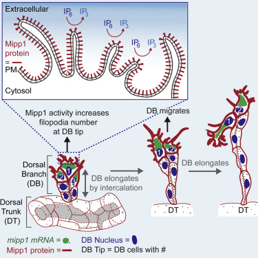


Cells 'reach out' to build trachea

Mipp1 protein helps cells sprout 'fingers' in order to grab nearby cells and pull them into production of new tissue structures in the fruit fly. Do some of our cells do the same?


Fruit fly windpipes are more like our human blood vessels than entryways to the lungs. According to Deborah Andrew PhD, professor of cell biology at the Johns Hopkins University School of Medicine: "Fruit flies don't have blood to bring oxygen to their cells. Instead, the tubes of the windpipe, or trachea, branch out repeatedly, getting thinner and thinner — like the tiny capillary blood vessels throughout our bodies — so that oxygen can diffuse directly from the trachea into nearby tissue."

To create this intricate network, fly embryo cells must sprout "fingers" and crawl into place. Now researchers at The Johns Hopkins University have discovered that a protein called Mipp1 is key to trachea cells' ability to grow such 'fingers.'

A summary of this research, which has implications for understanding normal and abnormal development in human and animal tissues, is published online in the journal Cell Reports.

Fruit flies are a research animal favorite among biologists because their genes and chemistry are relatively easy to manipulate. It also helps that they easily and quickly breed to improve their status as a model animal to study. As evolution highly conserves key biological events, what is learned from fruit flies may shed light on development in other species, including humans.

Andrew points out that the two major ducts of the embryonic fruit fly trachea run parallel to each other along the length of the embryo body. From these wide "dorsal trunks," several thinner branches split off and grow toward the top of the embryo where they meet and merge midline, forming a contiguous network.


Before and after elongating, the dorsal branches are just six interconnected cells. They start off stacked three high and wrapped around a thin tube connected to the dorsal trunk.

In order to elongate the tube, the cells at the top grow fingerlike structures called filopodia that reach out and pull the cells away from the dorsal trunk as the cells rearrange themselves to form a structure six cells high.


"A few years ago, we discovered that in developing fly embryos, the protein Mipp1 is controlled by a master regulator gene orchestrating all of tracheal development," says Yim Ling Cheng, Ph.D., a cell biology postdoctoral fellow at the Johns Hopkins University School of Medicine, and first author. Researchers had known that Mipp1 is an enzyme responsible for turning off chemical messenger molecules IP6 into IP3 ¬ by breaking off three of the phosphate groups. But, how is Mipp1 doing this.

By tracking Mipp1, they found the protein is located throughout the developing fruitfly, but soon becomes concentrated in the top pair of cells in the three-cell-high dorsal branches just before elongation. Those are the cells that grow filopodia. And, when there was too much Mipp1, the research team saw too many filopodia. Too little Mipp1 resulted in too few filopodia and branches that were slow to elongate.

Wondering if Mipp1's presence in the top cells was the cause or result of a cells' position, researchers genetically manipulated the flies so that dorsal branch cells turned on the Mipp1 gene at random.


They expected Mipp1 also to be found in the six positions of the trachial branch at random, but instead found Mipp1 in the top two cells at three times the concentratiion as in other cells. This suggests that Mipp1 makes cells more likely to climb to the top, where they are needed to elongate tracheal branches.

Further experimentation revealed Mipp1 decorates the outside edge — not the interior — of top cells of the tracheal branches, where it converts IP6 into IP3, but they wonder how exactly that influences finger growth. They hope to find out in their ongoing experiments.


Abstract Highlights
•Drosophila Mipp1 is expressed at high levels in leading cells of migrating trachea
•Mipp1 localizes to the plasma membrane and converts extracellular IP6 to IP3
•Mipp1 facilitates filopodia formation and cell rearrangement during tube elongation
•Mipp1 confers the same migration advantage to cells as highest level FGF signaling

Summary
Multiple Inositol Polyphosphate Phosphatase (Mipp), is a highly conserved but poorly understood histidine phosphatase, dephosphorylates higher-order IPs (IP4–IP6) to IP3. To gain insight into the biological roles of these enzymes, we have characterized Drosophila mipp1. Mipp1 is dynamically expressed in the embryonic trachea, specifically in the leading cells of migrating branches at late stages, where Mipp1 localizes to the plasma membrane and filopodia. FGF signaling activates mipp1 expression in these cells, where extensive filopodia form to drive migration and elongation by cell intercalation. We show that Mipp1 facilitates formation and/or stabilization of filopodia in leading cells through its extracellular activity. Mipp1 loss decreases filopodia number, whereas Mipp1 overexpression increases filopodia number in a phosphatase-activity-dependent manner.

Importantly, expression of Mipp1 gives cells a migratory advantage for the lead position in elongating tracheal branches. Altogether, these findings suggest that extracellular pools of inositol polyphosphates affect cell behavior during development.

This work was supported by grants from the National Institute of Dental and Craniofacial Research (RO1 DE012873, F31 DE021285).

Return to top of page

Dec 2, 2015   Fetal Timeline   Maternal Timeline   News   News Archive   


A schematic showing of the elongation of the developing fruit fly trachea
— and its "fingers."
Image Credit: Cell Press


Phospholid by Wikipedia