|
|||||||||||||||
|

CLICK ON weeks 0 - 40 and follow along every 2 weeks of fetal development
|
||||||||||||||||||||||||||||
|
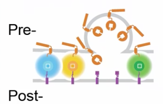
Neuroscientists now can read the mind of a fly Mapping the pattern of individual neural connections could provide insights into the computational processes that underlie the workings of our human brain. The study focused on three of the fruit fly's sensory systems. Researchers used fluorescent molecules of different colors to tag neurons in the brain to see which connections were active during a sensory experience that happened hours earlier. Synapses are points of communication where neurons exchange information. The fluorescent labeling technique is the first to allow scientists to identify individual synapses that are active during a complex behavior, such as avoiding heat. Better yet, the fluorescent signal persists for hours after the communication event, allowing researchers to study the brain's activity after the fact, under a microscope.
For example, by reading the fluorescent signals, researchers could tell if a fly had been in either heat or cold for 10 minutes an entire hour after the sensory event had happened. They also could see that exposure to the scent of a banana activated neural connections in the olfactory system that were different from those activated when the fly smelled jasmine. Details of the versatile technique, which could be used with other model systems for neuroscience study, are published in the journal Nature Communications. Gallio and team wanted to see brain activity of a fruit fly while it performed a complex behavior, but this cannot easily be achieved under a microscope. So, the scientists figured out a different approach using genetic engineering. Using the gene for a green fluorescent protein found in jellyfish, the authors derived three different colored markers that light up on synapse between active neurons. Additionally, the fluorescent signals can be read one to three hours after the neural action is over. "Different synapses are active during different behaviors, and we can see that in the same animal with our three distinct labels," said Gallio, the paper's corresponding author. The fluorescent green, yellow and blue signals enabled researchers to label different synapses to be activated by sensory experience in unique colors within the same animal. The fluorescent signals persisted and could later be viewed under a relatively simple microscope.
They exposed the flies to different sensory experiences, such as heat or light exposure and smelling bananas or jasmine, to see what was happening in their brains during each experience. To create labels, they had to split each fluorescent molecule in half, one half attached to the talking neuron and one half attached to the listening neuron. If those neurons "talked" to each other when a fly was exposed to a banana smell or to heat, the two halves of the molecule came together and lit up. This only happened at the site of active synaptic transmission.
This is a type of new technology discussed in President Obama's BRAIN Initiative (Brain Research Through Advancing Innovative Neurotechnologies). Such a tool will help research better understand how brain circuits process information. Abstract The paper is titled 'Dynamic labelling of neural connections in multiple colours by trans-synaptic fluorescence complementation.' In addition to Gallio, other authors of the paper include Emanuela E. Zaharieva, Patrick J. Kearney and Michael H. Alpert, of Northwestern; Lindsey J. Macpherson (first author) and Zeynep Turan, of Columbia University; and Tzu-Yang Lin and Chi-Hon Lee, of the Eunice Kennedy Shriver National Institute of Child Health and Human Development, National Institutes of Health. |
Dec 11, 2015 Fetal Timeline Maternal Timeline News News Archive
|
||||||||||||||||||||||||||||