|
Molecule stops brain stem cells from becoming tumors
Within natural aging of neural stem cells in the fruit fly, science has uncovered a molecular change. They have found a protein which is expressed (turned on) that blocks tumor formation.
The precisely timed changes in aging stem cells during development of the fly's central nervous system, could show us a molecular path to identify some cancers. If so, we could then target therapies based around those molecular changes, say University of Oregon (UO) scientists.
The research, detailed in the journal Current Biology, focused on the embryonic or larval stage of fruit fly (Drosophila) development. This is when stem cells generate most of the neurons forming the adult brain.
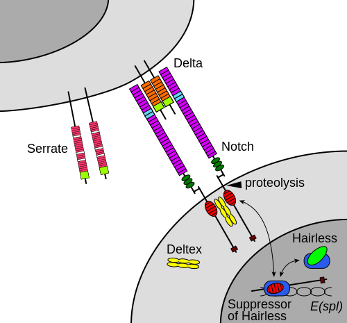
While rapidly dividing to produce new cells, brain stem cells rely on signals initiated by the protein Notch. The Notch signaling pathway plays an important role in cell to cell communication by regulating embryonic development. It is also a protein signaling system known to play a strong role in neural growth and differentiation, according to Tuft's University research in March 2015 (VisEmbryo).
Previous research has shown that when Notch signaling runs smoothly, stem cells make neurons that populate the adult central nervous system. But, too much Notch and stem cells over-proliferate — forming large tumors.
In humans, adult T-cell leukemia is tied to overactive Notch signaling.
"Stem cells have a really tough job, because they divide to make millions of neurons in our brain. When they don't divide enough, it results in microcephaly or other small brain diseases. But if they divide too much, they make tumors.
"They have to stay right on that boundary of dividing to make neurons but not dividing excessively and forming a tumor. They really walk a tightrope."
Chris Doe PhD, Professor of Biology, a Howard Hughes Medical Institute Investigator based at the University of Oregon (UO), and co-author.
In this new research, UO doctoral student Dylan Farnsworth observed that allowing stem cells to divide a few times, they age a bit and quit responding to Notch. At that point, stem cells cannot be pushed by high doses of Notch signaling to form tumors. He wondered what controlled Notch at this point.
Further observation led to the discovery of an age-related molecular change. Late in stem cell lives — about the same time they resist tumor formation — Drosophila stem cells begin expressing a transcription protein known as Eyeless (or Pax6 in humans). Eyeless appeared to block Notch signaling.
When the protein Eyeless is knocked out [turned off] in brain stem cells, Notch signaling overwhelms stem cells — which then form brain tumors.
Farnsworth: "If we can identify the stem cells that are relied upon during development, maybe we could find a way to use them later to re-create conditions that might be therapeutic."
Finding the role of Eyeless is a step in that direction, but it's possible that other molecules also are present which may inhibit tumor growth, Doe adds.
"This paper shows that Eyeless is important for winding down the lifespan of the stem cells that are giving rise to the adult brain. It's a stop signal that says it is time to cease responding to Notch signals."
Chris Doe PhD.
The system used to probe these cells was created purely for basic science research in Doe's lab. But now, it opens a new window to identify cells involved in the origin of cancers.
Tumors induced by Notch signaling in older stem cells were distinct from earlier forming masses. According to Doe, with more extensive research, the system could provide a roadmap to fine-tuning the timing of stem cell-based therapies and restart healthy activity in adult stem cells.
Abstract Highlights
•Aging INPs lose competence to respond to constitutively active Notch signaling
•The late temporal factor Eyeless blocks Notch-induced target gene expression
•Eyeless blocks Notch-induced INP tumor formation
Summary
Drosophila neural stem cells (neuroblasts) are a powerful model system for investigating stem cell self-renewal, specification of temporal identity, and progressive restriction in competence. Notch signaling is a conserved cue that is an important determinant of cell fate in many contexts across animal development; for example, mammalian T cell differentiation in the thymus and neuroblast specification in Drosophila are both regulated by Notch signaling. However, Notch also functions as a mitogen, and constitutive Notch signaling potentiates T cell leukemia as well as Drosophila neuroblast tumors. While the role of Notch signaling has been studied in these and other cell types, it remains unclear how stem cells and progenitors change competence to respond to Notch over time. Notch is required in type II neuroblasts for normal development of their transit amplifying progeny, intermediate neural progenitors (INPs). Here, we find that aging INPs lose competence to respond to constitutively active Notch signaling. Moreover, we show that reducing the levels of the old INP temporal transcription factor Eyeless/Pax6 allows Notch signaling to promote the de-differentiation of INP progeny into ectopic INPs, thereby creating a proliferative mass of ectopic progenitors in the brain. These findings provide a new system for studying progenitor competence and identify a novel role for the conserved transcription factor Eyeless/Pax6 in blocking Notch signaling during development.
Former UO doctoral student Omer Ali Bayraktar, now a postdoctoral fellow in the Department of Regenerative Medicine at the University of California, San Francisco, also was a co-author.
A grant from National Institutes of Health (T32 GM007413) to Farnsworth helped fund the research along with HHMI support to Doe.
Sources: Chris Doe, UO professor of biology, 541-346-4877, cdoe@uoregon.edu, and Dylan Farnsworth, doctoral student, Department of Biology, 541-954-5496, drf@uoregon.edu
Return to top of page
|