Welcome to The Visible Embryo
  o
 
The Visible Embryo Home
   
Google  
Home--- -History-----Bibliography-----Pregnancy Timeline-----Prescription Drugs in Pregnancy---- Pregnancy Calculator----Female Reproductive System----News----Contact
 
Welcome to The Visible Embryo, a comprehensive educational resource on human development from conception to birth.

The Visible Embryo provides visual references for changes in fetal development throughout pregnancy and can be navigated via fetal development or maternal changes.

The National Institutes of Child Health and Human Development awarded Phase I and Phase II Small Business Innovative Research Grants to develop The Visible Embryo. Initally designed to evaluate the internet as a teaching tool for first year medical students, The Visible Embryo is linked to over 600 educational institutions and is viewed by more than one million visitors each month.

Today, The Visible Embryo is linked to over 600 educational institutions and is viewed by more than 1 million visitors each month. The field of early embryology has grown to include the identification of the stem cell as not only critical to organogenesis in the embryo, but equally critical to organ function and repair in the adult human. The identification and understanding of genetic malfunction, inflammatory responses, and the progression in chronic disease, begins with a grounding in primary cellular and systemic functions manifested in the study of the early embryo.

WHO International Clinical Trials Registry Platform


The World Health Organization (WHO) has created a new Web site to help researchers, doctors and patients obtain reliable information on high-quality clinical trials. Now you can go to one website and search all registers to identify clinical trial research underway around the world!


Home

History

Bibliography

Pregnancy Timeline

Prescription Drug Effects on Pregnancy

Pregnancy Calculator

Female Reproductive System

News

Disclaimer: The Visible Embryo web site is provided for your general information only. The information contained on this site should not be treated as a substitute for medical, legal or other professional advice. Neither is The Visible Embryo responsible or liable for the contents of any websites of third parties which are listed on this site.


Content protected under a Creative Commons License.
No dirivative works may be made or used for commercial purposes.


Pregnancy Timeline by SemestersDevelopmental TimelineFertilizationFirst TrimesterSecond TrimesterThird TrimesterFirst Thin Layer of Skin AppearsEnd of Embryonic PeriodEnd of Embryonic PeriodFemale Reproductive SystemBeginning Cerebral HemispheresA Four Chambered HeartFirst Detectable Brain WavesThe Appearance of SomitesBasic Brain Structure in PlaceHeartbeat can be detectedHeartbeat can be detectedFinger and toe prints appearFinger and toe prints appearFetal sexual organs visibleBrown fat surrounds lymphatic systemBone marrow starts making blood cellsBone marrow starts making blood cellsInner Ear Bones HardenSensory brain waves begin to activateSensory brain waves begin to activateFetal liver is producing blood cellsBrain convolutions beginBrain convolutions beginImmune system beginningWhite fat begins to be madeHead may position into pelvisWhite fat begins to be madePeriod of rapid brain growthFull TermHead may position into pelvisImmune system beginningLungs begin to produce surfactant
CLICK ON weeks 0 - 40 and follow along every 2 weeks of fetal development


Messenger RNA may fine tune protein production

Long thought of as a simple link between DNA and protein, new research at The Rockefeller University suggests messenger RNA is up to something unexpected.


By uncovering widespread differences in the expression of parts making up the mRNA molecule — something assumed not to happen — scientists think they have found intriguing patterns suggesting functions for regions within mRNA molecules competely unexpected.

"The lopsided ratios we found in two parts of mRNA — one that carries the code for a protein and one that doesn't — do not appear to be random," says senior author Mary Hynes, a research associate professor. "We suspect some of these skewed ratios may be regulating protein production, particularly during embryonic development, but also in adults."

Reported December 16 in Neuron, the research results focus on regions within messenger RNA that imparted different cell responses.


RNA is transcribed directly from a gene, then is translated into protein.

After being transcribed, each mRNA molecule possesses a coding sequence in it's middle, which a protein reads for instructions.

This coding sequence is flanked on either end by two other sections that do not make protein, the 3' untranslated region (UTR) and the 5' untranslated region (UTR), each name refers to the sugar backbone of the molecule.


The 5' untranslated region (5′ UTR) is also known as a Leader Sequence or Leader RNA. It is the region of mRNA directly upstream giving the signal for the beginning of transcription. It specifies the first amino acid in protein synthesis and so is important for regulating transcription.

It had long been assumed all three sections stick together as a single mRNA molecule to make transcribing RNA work.

However, using a technique developed at Rockefeller University called translating ribosome affinity purification or TRAP, the Hynes' team isolated purified mRNA from dopamine neurons in mouse embryos. TRAP is a new technique (2014) that shows the entire cell population of mRNA, and reduces it's complexity. Its a view that helps observers understand cell function on the molecular level.


With this technique, the scientists made a surprising observation: although dopamine neuron genes show abundant expression of the 3' untranslated region (UTR) sequence, there was little or no expression of the 5' untranslated region (UTR) or coding region.

This was seen for Sox11 and Sox12 genes which are known to help determine cell fate during development.

This contradicts common thinking that once an mRNA is transcribed from a gene and a protein expressed, the 5' and 3'UTR coding regions continue to act as one unit. So when protein is produced, would signal mRNA to be degraded and recycled.


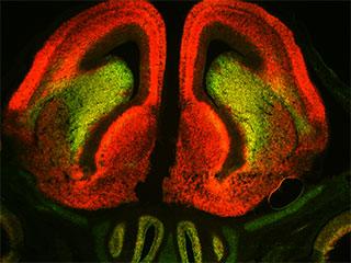
An earlier study noted similar disparities, but did not explore the biological implications and that study's data did not get widespread attention. Hynes and her colleagues took the next step, asking: Why would a cell make abundant levels of 3'UTR sequence with no 5'UTR coding sequence, since protein cannot be made without the coding sequence?

To verify their findings and understand if this was restricted to dopamine neurons, development, or just the nervous system, the scientists used green probes to mark 5'UTR coding sequences and red to mark the 3'UTRs of 19 genes in embryonic and adult tissue.

"Based on prior understanding, it was expected that every cell in the tissue should show up as either yellow, when both are expressed, or black, when neither are," Hynes says. "But to our surprise, when we examined Sox11 mRNA in the brain we found many neurons that were red, or expressing mostly UTR, as well as many that were green, or expressing mostly coding sequences."

They went on to show this was true for every gene examined and that different levels of expression of UTR and coding sequences occurs throughout the embryo, the adult, and outside of the nervous system.


Even widely expressed genes such as beta actin, a protein needed for cell movement and structure, show differences in UTR and coding sequence amounts.

Taking all protein expression into account, they found the higher the ratio of 3' UTR to coding sequence, the lower the level of a protein.

This suggests high levels of 3' UTR might be involved in turning the dial down on protein production.


However, it's not clear how this might happen, Hynes adds.

Next, they focused back on the developing dopamine neurons and the nine thousand genes they found active in them.


With help from the New York Genome Center, the team compared the biological functions of genes with a high UTR-to-coding sequence ratio against those with similar ratios.

Many of the high UTR genes turned out to play roles specific to development, while the genes with similar ratios were more often involved in generic cell processes.


Adds Hynes: "During development a neuron may need to express a certain gene, but only a particular amount. Either too much or too little might be harmful and lead to irreversible changes. So, we think this could be a mechanism for finely titrating [adjusting the balance] of proteins levels in an active gene."


Hynes believes that because RNA expression can rely on sequences within the UTRs, the coding section, or both, potentially essential information may be lost.


Hynes: "Going forward, I think that when an RNA sequencing experiment suggests that a gene is highly expressed, researchers should take a closer look at the relative levels of these two other components to get a more accurate picture of what is being expressed."

Abstract Highlights
•mRNA coding (CDS) and cognate 3′ UTR regions show widespread unbalanced expression
•A given gene can show a broad range of 3′ UTR to CDS expression ratios across neurons/cells
•Ratios are spatially graded and change with age but are consistent across embryos
•A high 3′ UTR-to-CDS ratio may predict lower protein level

Summary
Mature messenger RNAs (mRNAs) consist of coding sequence (CDS) and 5′ and 3′ UTRs, typically expected to show similar abundance within a given neuron. Examining mRNA from defined neurons, we unexpectedly show extremely common unbalanced expression of cognate 3′ UTR and CDS sequences; many genes show high 3′ UTR relative to CDS, others show high CDS to 3′ UTR. In situ hybridization (19 of 19 genes) shows a broad range of 3′ UTR-to-CDS expression ratios across neurons and tissues. Ratios may be spatially graded or change with developmental age but are consistent across animals. Further, for two genes examined, a 3′ UTR-to-CDS ratio above a particular threshold in any given neuron correlated with reduced or undetectable protein expression. Our findings raise questions about the role of isolated 3′ UTR sequences in regulation of protein expression and highlight the importance of separately examining 3′ UTR and CDS sequences in gene expression analyses.

Return to top of page

Dec 25, 2015   Fetal Timeline   Maternal Timeline   News   News Archive   


When 3' UTR sequences of Sox 11 mRNA in embryonic mouse brain (above) were marked in red, coding sequences appeared in green. Common thinking suggested all Sox 11-expressing cells would appear yellow, indicating both components were present in even amounts.
Clearly 3' UTR sequences cells are red and cells higher in Sox 11 mRNA appear in green.
Image Credit: Lab. Neural Specification and Development/Rockefeller University


Phospholid by Wikipedia