Welcome to The Visible Embryo
  o
 
The Visible Embryo Home
   
Google  
Home--- -History-----Bibliography-----Pregnancy Timeline-----Prescription Drugs in Pregnancy---- Pregnancy Calculator----Female Reproductive System----News----Contact
 
WHO International Clinical Trials Registry Platform


The World Health Organization (WHO) has a Web site to help researchers, doctors and patients obtain information on clinical trials. Now you can search all such registers to identify clinical trial research around the world!


Home

History

Bibliography

Pregnancy Timeline

Prescription Drug Effects on Pregnancy

Pregnancy Calculator

Female Reproductive System

News

Disclaimer: The Visible Embryo web site is provided for your general information only. The information contained on this site should not be treated as a substitute for medical, legal or other professional advice. Neither is The Visible Embryo responsible or liable for the contents of any websites of third parties which are listed on this site.


Content protected under a Creative Commons License.
No dirivative works may be made or used for commercial purposes.


Pregnancy Timeline by SemestersDevelopmental TimelineFertilizationFirst TrimesterSecond TrimesterThird TrimesterFirst Thin Layer of Skin AppearsEnd of Embryonic PeriodEnd of Embryonic PeriodFemale Reproductive SystemBeginning Cerebral HemispheresA Four Chambered HeartFirst Detectable Brain WavesThe Appearance of SomitesBasic Brain Structure in PlaceHeartbeat can be detectedHeartbeat can be detectedFinger and toe prints appearFinger and toe prints appearFetal sexual organs visibleBrown fat surrounds lymphatic systemBone marrow starts making blood cellsBone marrow starts making blood cellsInner Ear Bones HardenSensory brain waves begin to activateSensory brain waves begin to activateFetal liver is producing blood cellsBrain convolutions beginBrain convolutions beginImmune system beginningWhite fat begins to be madeHead may position into pelvisWhite fat begins to be madePeriod of rapid brain growthFull TermHead may position into pelvisImmune system beginningLungs begin to produce surfactant
CLICK ON weeks 0 - 40 and follow along every 2 weeks of fetal development


Potential to treat hypertrophic cardiomyopathy

More than 15 years ago, researchers discovered the precise malfunction of a specific protein in the heart that leads to hypertrophic cardiomyopathy (HCM), a common culprit in the sudden death of young athletes.


Now, a team of scientists led by David Warshaw, has used their earlier findings to develop a possible treatment to prevent HCM, an inherited disease which can cause the heart to thicken and stop effectively pumping blood — ending in heart failure. Warshaw is professor and chair of molecular physiology and biophysics at the University of Vermont (UVM) College of Medicine. He wrote about the significance of this potential therapy for a "Perspectives" column in the February 5, 2016 issue of the journal Science.

"This may offer a generalized approach to solving hypertrophic cardiomyopathy," says David Warshaw PhD, who is also an investigator in the Cardiovascular Research Institute of Vermont at UVM. "I think it's extremely promising."


HCM can result from different mutations of many proteins in the heart. One of those proteins, myosin, acts as a tiny molecular motor in every heart muscle cell. It pulls on and releases on a rope-like protein, actin, in order to make the heart muscle contract and relax as it pumps blood.


A mutation of myosin can "alter the motor's power-generating capacity" and make the heart work improperly, which in turn causes the heart to enlarge, Warshaw says.

For many years, scientists assumed that the mutation caused the myosin to lose its motoring power, throwing off the whole heart engine. But in a study Warshaw published in 2000 in Circulation Research, he and colleagues found that the problem wasn't diminished power in the myosin, it was too much power in myosin with this mutation.

"By analogy, placing the engine of an Indy race car (i.e., mutant myosin) in a stock car chassis (i.e., the heart's connective tissue matrix) could lead to internal stress and structural damage," Warshaw writes in his "Perspectives" article. "For the heart, this amounts to inducing cardiac fibrosis and muscle cell disarray characteristic of HCM patients."


The team of scientists who found a way to address this problem - which they report in the February 5, 2016 issue of Science - are from Harvard Medical School, Stanford University School of Medicine, University of Colorado, and MyoKardia Inc. in San Francisco, a biotechnology company formed to develop such treatments.

Using mice bred with the mutation, the team tested a molecular inhibitor that dials down the power of the myosin motor to a more normal level. As early as eight weeks old, mice in the study that received the drug containing the molecule did amazingly well and the HCM was prevented from surfacing.

"When they gave the drug to a young mouse with the mutation, the mouse's heart developed normally," Warshaw adds.


Because HCM runs in families, an infant who tests positive for the genetic mutation could receive the treatment and stave off the disease, believes Warshaw. However, development of a human drug would require more extensive testing and there are many questions remaining.


Nonetheless, Warshaw sees great potential. In previous studies, he found mutations to other heart proteins also increased the heart muscle, leading to HCM. Warshaw believes the same molecule could be used on the myosin motor to compensate and block HCM in all related cases.

Abstract: Science Perspectives: "A small-molecule inhibitor of sarcomere contractility suppresses hypertrophic cardiomyopathy in mice"
Hypertrophic cardiomyopathy (HCM) is an inherited disease of heart muscle that can be caused by mutations in sarcomere proteins. Clinical diagnosis depends on an abnormal thickening of the heart, but the earliest signs of disease are hyperdynamic contraction and impaired relaxation. Whereas some in vitro studies of power generation by mutant and wild-type sarcomere proteins are consistent with mutant sarcomeres exhibiting enhanced contractile power, others are not. We identified a small molecule, MYK-461, that reduces contractility by decreasing the adenosine triphosphatase activity of the cardiac myosin heavy chain. Here we demonstrate that early, chronic administration of MYK-461 suppresses the development of ventricular hypertrophy, cardiomyocyte disarray, and myocardial fibrosis and attenuates hypertrophic and profibrotic gene expression in mice harboring heterozygous human mutations in the myosin heavy chain. These data indicate that hyperdynamic contraction is essential for HCM pathobiology and that inhibitors of sarcomere contraction may be a valuable therapeutic approach for HCM.

Summary: Science Magazine: "Throttling back the heart's molecular motor"
A young athlete collapses and dies during competition. Autopsy reveals an enlarged heart with thickened walls in which the cardiac muscle cells are in disarray and surrounded by fibrotic tissue. Until 1990, the cause of such sudden death was unknown. This devastating condition, called familial hypertrophic cardiomyopathy (HCM), was eventually linked to a mutation in myosin (1), the heart's molecular motor. Today, more than 300 separate HCM-causing mutations have been identified throughout the myosin molecule. On page 617 of this issue, Green et al. (2) describe a small molecule that binds to myosin and inhibits its activity, delaying the onset and progression of the disease in a mouse model. The study offers hope that a “simple” remedy for HCM may be possible.

Return to top of page

Feb 17, 2016   Fetal Timeline   Maternal Timeline   News   News Archive   

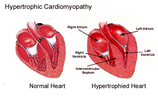
Scientists have identified a small molecule, MYK-461, that reduces contractility of the heart muscle
by decreasing the adenosine triphosphatase activity of the cardiac myosin heavy chain. Here we
demonstrate that early, chronic administration of MYK-461 suppresses the development of
ventricular hypertrophy, cardiomyocyte disarray, and myocardial fibrosis.
Image Credit:
Nature Communications


Phospholid by Wikipedia