Welcome to The Visible Embryo
  o
 
The Visible Embryo Home
   
Google  
Home--- -History-----Bibliography-----Pregnancy Timeline-----Prescription Drugs in Pregnancy---- Pregnancy Calculator----Female Reproductive System----News----Contact
 
WHO International Clinical Trials Registry Platform


The World Health Organization (WHO) has a Web site to help researchers, doctors and patients obtain information on clinical trials. Now you can search all such registers to identify clinical trial research around the world!


Home

History

Bibliography

Pregnancy Timeline

Prescription Drug Effects on Pregnancy

Pregnancy Calculator

Female Reproductive System

News

Disclaimer: The Visible Embryo web site is provided for your general information only. The information contained on this site should not be treated as a substitute for medical, legal or other professional advice. Neither is The Visible Embryo responsible or liable for the contents of any websites of third parties which are listed on this site.


Content protected under a Creative Commons License.
No dirivative works may be made or used for commercial purposes.


Pregnancy Timeline by SemestersDevelopmental TimelineFertilizationFirst TrimesterSecond TrimesterThird TrimesterFirst Thin Layer of Skin AppearsEnd of Embryonic PeriodEnd of Embryonic PeriodFemale Reproductive SystemBeginning Cerebral HemispheresA Four Chambered HeartFirst Detectable Brain WavesThe Appearance of SomitesBasic Brain Structure in PlaceHeartbeat can be detectedHeartbeat can be detectedFinger and toe prints appearFinger and toe prints appearFetal sexual organs visibleBrown fat surrounds lymphatic systemBone marrow starts making blood cellsBone marrow starts making blood cellsInner Ear Bones HardenSensory brain waves begin to activateSensory brain waves begin to activateFetal liver is producing blood cellsBrain convolutions beginBrain convolutions beginImmune system beginningWhite fat begins to be madeHead may position into pelvisWhite fat begins to be madePeriod of rapid brain growthFull TermHead may position into pelvisImmune system beginningLungs begin to produce surfactant
CLICK ON weeks 0 - 40 and follow along every 2 weeks of fetal development


Zika virus breakthrough

Florida State University research has found that the Zika virus creates birth defects by specifically targeting and stunting the growth of developing brain cells.


This is the first major finding by scientists to verify that developing brain cells are the critical target of the virus. Hengli Tang, professor of biological science at Florida State University (FSU), is a lead author of the study published March 4 in the journal Cell Stem Cell.

"We aim to fill the knowledge gap between infection and neurological defects," Tang adds. "This research is the very first step. Now we can be studying the virus in the right cell type, screening drugs on the right cell type and studying the biology of the right cell type."

Though the Zika virus was discovered in 1947, there was very little known about how it worked or its health implications, especially among pregnant women. Anecdotal evidence has now suggested its' link to microcephaly, a condition in which a child is born with an abnormally small head with incomplete brain development.


Tang, along with colleagues at Johns Hopkins and Emory Universities, found the Zika virus directly targets Human Neural Progenitor Cells (hNPCs) — in as little as 3 days following exposure. Infected cells reproduce the virus, interrupting cell function and growth. Some cells die immediately upon infection.


"Potentially, this could explain why there is a link to microcephaly, but there is a lot more work needed to show direct cause," adds Guo-Li Ming, professor of neurology at Johns Hopkins University.

The research is taking a remarkably swift path. To protect public health, research worldwide is working around the clock to find how the virus works and its potential targets.


According to the World Health Organization, 48 countries report transmission of the Zika virus.


About a month ago, a research team led by Johns Hopkins University Neurology Professors Hongjun Song and Guo-li Ming, brought neural stem cells to FSU in the United States, where Tang and his graduate students infected them with Zika in order to monitor gene expression, or how genes are turned on and off, by the virus. A few weeks later, the cells were taken to Emory University for more analysis of change.

Tang has in the past received funding from the National Institutes of Health to study Dengue virus, similar to Zika, and runs a lab equipped to handle and study virus. Song and Ming are fellow graduate students with Tang, and experts on neural cells believed to be targets of Zika.

The three labs collaborate as well as separately tackle, various aspects of Zika. Tang is investigating how Zika enters the cell and disrupts normal cell processes. Ming creates 3D brain models to examine any link between neural progenitor cells and microcephaly. And Song investigates why the virus is targeting neural progenitor cells as opposed to other cell types.


Many questions still remain about the virus, but this discovery is the pivotal first step.


"It's significant because we're literally the first people in the world to know this, to know that this virus can infect these very important cells and interfere with their function," Tang explains. "Research is rewarding in general, but when you have something this timely and this clinically relevant, it's extra satisfying. We are helping people in the long run."

From the research:
Flaviviruses tend to have broad cellular tropisms and multiple factors contribute to pathogenic outcomes, including specific cellular response and tissue accessibility. Dengue virus infects cells of several lineages and hematopoietic cells play an essential role in the associated pathogenesis (Pham et al., 2012). West Nile virus infects epithelial cells of multiple tissues and can be neuroinvasive (Suthar et al., 2013). We note that ZIKV also infects other human cell types, including skin cells and fibroblasts (Hamel et al., 2015), and it remains unknown how ZIKV may gain access to the fetal brain (Mlakar et al., 2016). The capacity of ZIKV to infect hNPCs and attenuate their growth underscores the urgent need for more research into the role of these cells in putative ZIKV-related neuropathology. The finding that ZIKV also infects immature neurons raises critical questions about pathological effects on neurons and other neural cell types in the brain, as well as potential long-term consequences. Intriguingly, an early animal study showed ZIKV infection of neurons and astrocytes in mice and observed enlarged astrocytes (Bell et al., 1971). Our study also raises the question of whether ZIKV infects neural stem cells in adult humans (Bond et al., 2015).

Other authors on the paper are Florida State University researcher Ruth Didier and graduate students Christy Hammack, Sarah Ogden and Emily Lee; Zhexing Wen, Xuyu Qian and Kimberly Christian from Johns Hopkins University; and Yujing Li, Bing Ya, Feiran Zhang and Peng Jin from Emory University.

The research was funded by Florida State University, the Maryland Stem Cell Research Fund and the National Institutes of Health.
Return to top of page

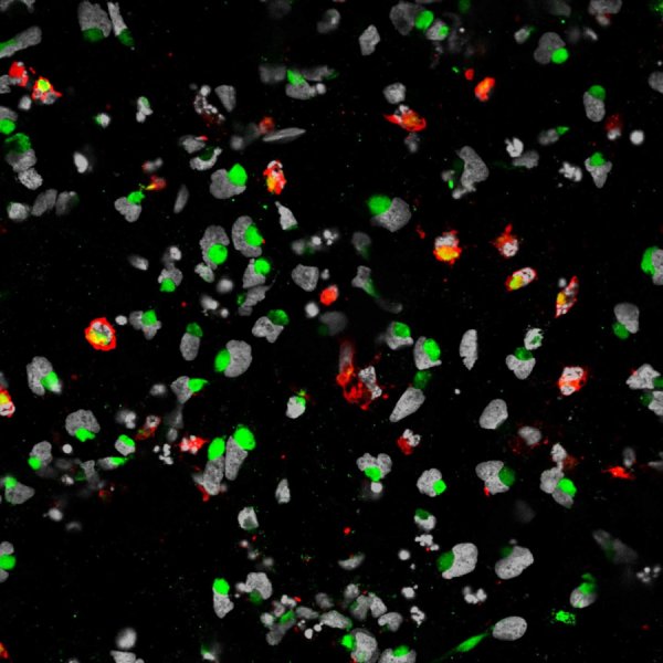
Mar 14, 2016   Fetal Timeline   Maternal Timeline   News   News Archive   

ZIKV (The Zika virus) infects a type of neural stem cell that gives rise to the brain's cerebral cortex.
In laboratory dishes, these stem cells were found to be havens for viral reproduction, resulting in
cell death and/or disruption of cell growth. While this study does not prove the direct link between
Zika and microcephaly, it does pinpoint where the virus may be doing the most damage.

Image Credit: Guo-Li Ming Laboratory, Johns Hopkins University, USA


Phospholid by Wikipedia