Welcome to The Visible Embryo
  o
 
The Visible Embryo Home
   
Google  
Home--- -History-----Bibliography-----Pregnancy Timeline-----Prescription Drugs in Pregnancy---- Pregnancy Calculator----Female Reproductive System----News----Contact
 
WHO International Clinical Trials Registry Platform


The World Health Organization (WHO) has a Web site to help researchers, doctors and patients obtain information on clinical trials. Now you can search all such registers to identify clinical trial research around the world!


Home

History

Bibliography

Pregnancy Timeline

Prescription Drug Effects on Pregnancy

Pregnancy Calculator

Female Reproductive System

News

Disclaimer: The Visible Embryo web site is provided for your general information only. The information contained on this site should not be treated as a substitute for medical, legal or other professional advice. Neither is The Visible Embryo responsible or liable for the contents of any websites of third parties which are listed on this site.


Content protected under a Creative Commons License.
No dirivative works may be made or used for commercial purposes.


Pregnancy Timeline by SemestersDevelopmental TimelineFertilizationFirst TrimesterSecond TrimesterThird TrimesterFirst Thin Layer of Skin AppearsEnd of Embryonic PeriodEnd of Embryonic PeriodFemale Reproductive SystemBeginning Cerebral HemispheresA Four Chambered HeartFirst Detectable Brain WavesThe Appearance of SomitesBasic Brain Structure in PlaceHeartbeat can be detectedHeartbeat can be detectedFinger and toe prints appearFinger and toe prints appearFetal sexual organs visibleBrown fat surrounds lymphatic systemBone marrow starts making blood cellsBone marrow starts making blood cellsInner Ear Bones HardenSensory brain waves begin to activateSensory brain waves begin to activateFetal liver is producing blood cellsBrain convolutions beginBrain convolutions beginImmune system beginningWhite fat begins to be madeHead may position into pelvisWhite fat begins to be madePeriod of rapid brain growthFull TermHead may position into pelvisImmune system beginningLungs begin to produce surfactant
CLICK ON weeks 0 - 40 and follow along every 2 weeks of fetal development


Mom's bacterial infection can change fetal brain

St. Jude Children's Research Hospital discovers how pieces of bacteria cross the placenta and alter fetal brain anatomy affecting cognitive function after birth.


The study appears today in the journal Cell Host & Microbe.

Findings in the mouse as a model animal for the human brain, provide a possible mechanism which might underlie maternal bacterial infections in pregnancy with an increase in risk of autism and other cognitive problems in children.


The research raises the question "Which class of antibiotics could be used to treat such infections."


"The finding was unexpected, pneumococcal infections in children and adults can lead to meningitis and death of neurons," said the study's corresponding author Elaine Tuomanen, MD, and chair of the St. Jude Department of Infectious Diseases. "This study in a mouse model of bacterial infection found that prenatally the opposite is true. Evidence suggests maternal infections cause a signaling event that leads to proliferation and reorganization of neurons in a developing brain, which becomes defective in some way — maybe due to overcrowding."

Researchers revealed for the first time that pieces of the bacterial cell wall cross through the maternal placenta and travel into the fetal brain. This triggers proliferation of immature neurons. Evidence suggests the proliferation of neurons is sparked by a previously unrecognized pathway involving the immune system and a protein which regulates gene expression (or whether a gene is turned on or not turned on).


A 50 percent increase in neuron proliferation occurred in a region of the developing brain cortex. The cortex is responsible for thought, action and other higher cognitive function.


Investigators saw that mice exposed to pieces of bacterial cell wall early in fetal development, performed below average on tests measuring memory and cognitive function in mice after birth. They also found evidence that treatment of moms using the antibiotic ampicillin, which destroys bacteria and sends pieces of the bacterial cell wall into the bloodstream, still led to neuronal increase.

Tuomanen said the results raise questions about which class of antibiotics should be used to treat bacterial infections in pregnancy.


"This study suggests widely used antibiotics like ampicillin — that cause bacteria to burst cell walls — may lead to changes in the developing brain," she said. "Such changes did not occur in mice treated with antibiotics like clindamycin, which kills without releasing cell wall material.

"Additional studies are needed to understand the long-term impact of different classes of antibiotics on pregnancy outcomes," Tuomanen said.


Working with mouse cells grown in the lab, researchers found pneumococcal cell wall pieces bind to the protein platelet activating factor receptor (PAFr) to transport across the placental wall and enter the fetal brain.


Within hours of being in the fetal brain, instead of triggering inflammation and cell death, bacterial cells switched on the protein FoxG1. FoxG1 drives neural proliferation.

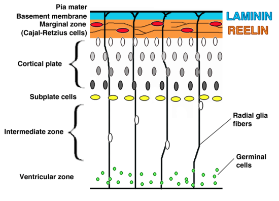
This occurred early in pregnancy and deep in the brain region called the ventricular zone. Within days, researchers documented a 50 percent increase in neurons in the cortical plate, part of the cortex.


Researchers also saw that some components of the immune system drove proliferation rather than inflammation.

A protein called toll-like receptor 2 (TLR2) typically helps cells recognize and defend against bacteria. Evidence from the study suggests TLR2 partnered with the protein TLR6 to drive proliferation instead.

Without TLR2, immature fetal neurons did not proliferate.

Researchers also reported that proliferation did not occur in fetal mice that lacked either PAFr or TLR2, suggesting that both proteins play a role in the brain's response to bacterial cell wall.


"Additional research is needed to understand how bacterial cell wall induces proliferation via this newly identified path linking the innate immune receptor TLR2 with the transcription factor FoxG1 — to drive neural proliferation in the fetus. This mechanism might lead to new strategies to repair or replacement of neurons lost to illness and injury," adds Dr. Tuomane .

Abstract Highlights
•Bacterial cell wall (CW) traverses the mouse placenta and is detected in the fetal brain
•CW induces transcription factor FoxG1 in the fetal cortex leading to neuroproliferation
•FoxG1 induction and neuroproliferation require TLR2 in vivo and in vitro
•CW exposure in utero results in abnormal postnatal cognitive behavior

Summary
Maternal infection during pregnancy is associated with adverse outcomes for the fetus, including postnatal cognitive disorders. However, the underlying mechanisms are obscure. We find that bacterial cell wall peptidoglycan (CW), a universal PAMP for TLR2, traverses the murine placenta into the developing fetal brain. In contrast to adults, CW-exposed fetal brains did not show any signs of inflammation or neuronal death. Instead, the neuronal transcription factor FoxG1 was induced, and neuroproliferation leading to a 50% greater density of neurons in the cortical plate was observed. Bacterial infection of pregnant dams, followed by antibiotic treatment, which releases CW, yielded the same result. Neuroproliferation required TLR2 and was recapitulated in vitro with fetal neuronal precursor cells and TLR2/6, but not TLR2/1, ligands. The fetal neuroproliferative response correlated with abnormal cognitive behavior in CW-exposed pups following birth. Thus, the bacterial CW-TLR2 signaling axis affects fetal neurodevelopment and may underlie postnatal cognitive disorders.

Beth Mann, of St. Jude, and Jessica Humann, Ph.D., formerly of St. Jude and now of Florida Agricultural and Mechanical University, Tallahassee, Fla., are the first authors. The other authors are Geli Gao, Philip Moresco, Joseph Ramahi, Lip Nam Loh, Arden Farr and Richard Smeyne, all of St. Jude; and Yunming Hu, Kelly Durick-Eder and Sophie Fillon, all formerly of St. Jude.

The research was funded in part by grants (CA02176535, CA23944, R0127913) from the National Institutes of Health, and ALSAC.

Return to top of page

Mar 15, 2016   Fetal Timeline   Maternal Timeline   News   News Archive   


Brain cortex layers migrate from the ventricular zone (GREEN DOTS)
through the subplate to become the cognitive region of the brain.
Image Credit: Wikipedia


Phospholid by Wikipedia