Welcome to The Visible Embryo
  o
 
The Visible Embryo Home
   
Google  
Home--- -History-----Bibliography-----Pregnancy Timeline-----Prescription Drugs in Pregnancy---- Pregnancy Calculator----Female Reproductive System----News----Contact
 
WHO International Clinical Trials Registry Platform


The World Health Organization (WHO) has a Web site to help researchers, doctors and patients obtain information on clinical trials. Now you can search all such registers to identify clinical trial research around the world!


Home

History

Bibliography

Pregnancy Timeline

Prescription Drug Effects on Pregnancy

Pregnancy Calculator

Female Reproductive System

News

Disclaimer: The Visible Embryo web site is provided for your general information only. The information contained on this site should not be treated as a substitute for medical, legal or other professional advice. Neither is The Visible Embryo responsible or liable for the contents of any websites of third parties which are listed on this site.


Content protected under a Creative Commons License.
No dirivative works may be made or used for commercial purposes.


Pregnancy Timeline by SemestersDevelopmental TimelineFertilizationFirst TrimesterSecond TrimesterThird TrimesterFirst Thin Layer of Skin AppearsEnd of Embryonic PeriodEnd of Embryonic PeriodFemale Reproductive SystemBeginning Cerebral HemispheresA Four Chambered HeartFirst Detectable Brain WavesThe Appearance of SomitesBasic Brain Structure in PlaceHeartbeat can be detectedHeartbeat can be detectedFinger and toe prints appearFinger and toe prints appearFetal sexual organs visibleBrown fat surrounds lymphatic systemBone marrow starts making blood cellsBone marrow starts making blood cellsInner Ear Bones HardenSensory brain waves begin to activateSensory brain waves begin to activateFetal liver is producing blood cellsBrain convolutions beginBrain convolutions beginImmune system beginningWhite fat begins to be madeHead may position into pelvisWhite fat begins to be madePeriod of rapid brain growthFull TermHead may position into pelvisImmune system beginningLungs begin to produce surfactant
CLICK ON weeks 0 - 40 and follow along every 2 weeks of fetal development


Genes can cause neurological diseases

Spinocerebellar ataxia (SCA) is a genetic disease that causes wasting away of the cerebellum — the portion of our brain responsible for controlling voluntary muscle movement, like walking, speaking, even the direction our eyes move.


Currently, SCA has no cure or treatment and genetic mutations responsible for about 30 percent of cases are still unidentified. However, two different families with inherited SCA looking for treatment and unable to be helped, had gene samples sent to Hiroshima University to begin the process of identifying their new mutation.

After sequencing the genes of family members with SCA, a research team — led by Professor Hideshi Kawakami MD PhD, from the Department of Epidemiology at Hiroshima University — began statistical analysis comparing family members' DNA to unrelated people without SCA. The analysis identified which gene variation SCA family members shared that is not found in healthy people.

The research appears in the journal Molecular Brain.


The gene responsible for causing both families' SCA is located on Chromosome 17. A gene called CACNA1G which encodes for the protein Cav3.1, a gateway, or ion channel, inside nerve cells which conducts calcium into cells of the body.

Cav3.1 controls the flow of Calcium ions into nerves after receiving an electrical impulse from the brain. But, it had never been linked to SCA.

Changing a single letter in the DNA sequence on CACNA1G switches a single amino acid in the chain of 2,377 amino acids which connect together to build the Cav3.1 protein.


Researchers performed more experiments in culture to examine how the mutated Cav3.1 channel behaves and found this mutation makes calcium channels open at a lower threshold than in healthy cells.

"In the future, a drug modifying this channel may cure patients," suggests Prof. Kawakami.

Skin cells from one patient were used in generate pluripotent stem cells, allowing for a patient's neurons to be grown in the laboratory. These newly grown neurons showed no obvious physical deformities, which might fit with the normal progression of SCA. Depending on which SCA mutation a patient has, some patients may not experience symptoms until they are middle-aged.

Prof. Kawakami: "We might need some age-related factors to reproduce life-like cell behavior," so research will continue using these lab dish neurons to study Cav3.1 under more life-like conditions and in greater detail in the future.

T-type calcium channels are low-voltage. They open during membrane depolarization, mediating the amount of calcium flowing into cells. After being given the depolarizing signal, the amount of calcium flowing into a cell induces numerous physiological responses.


Within cardiac and smooth muscle cells, contraction of cell walls responds to increased or decreased calcium concentrations.


Calcium channels in the 1970s became distinguished in cells of the central nervous system as either Transient Opening calcium channels (T-type calcium channels) or the more well-known Long-Lasting calcium channels (L-type calcium channels).

T-type calcium
channels became known for their ability to activate a negative membrane response and for being unresponsive to calcium antagonist drugs.

These two distinct calcium channels are located in the brain, peripheral nervous system, heart, smooth muscle, bone, and endocrine system.


The distinct structure of T-type calcium allows for the conduction of calcium via a primary α1 subunit — or pore that channels calcium flow.


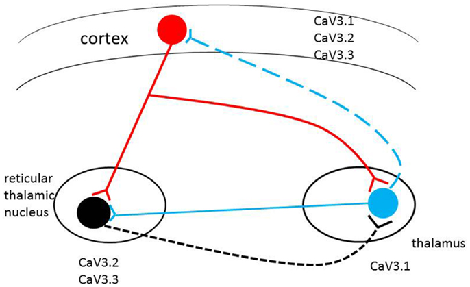
Through this pore, T-type calcium channels allow for the continuous rhythmic bursts of calcium opening valves in the heart, as well as for relaying rapid signal transmissions from the thalamus to numerous other locations throughout our body. And, as there is pharmacological evidence supporting these channels as key players in epilepsy, diabetes, and several forms of cancer, research continuously looks to create drugs to regulate their influence when irregular or missing.

Abstract Results (Open Article Online)
In this study, we analyzed a Japanese family with autosomal dominant SCA using linkage analysis and exome sequencing, and identified CACNA1G, which encodes the calcium channel CaV3.1, as a new causative gene. The same mutation was also found in another family with SCA. Although most patients exhibited the pure form of cerebellar ataxia, two patients showed prominent resting tremor in addition to ataxia. CaV3.1 is classified as a low-threshold voltage-dependent calcium channel (T-type) and is expressed abundantly in the central nervous system, including the cerebellum. The mutation p.Arg1715His, identified in this study, was found to be located at S4 of repeat IV, the voltage sensor of the CaV3.1. Electrophysiological analyses revealed that the membrane potential dependency of the mutant CaV3.1 transfected into HEK293T cells shifted toward a positive potential. We established induced pluripotent stem cells (iPSCs) from fibroblasts of the patient, and to our knowledge, this is the first report of successful differentiation from the patient-derived iPSCs into Purkinje cells. There was no significant difference in the differentiation status between control- and patient-derived iPSCs.

Conclusions
To date, several channel genes have been reported as causative genes for SCA. Our findings provide important insights into the pathogenesis of SCA as a channelopathy.
Return to top of page

Mar 17, 2016   Fetal Timeline   Maternal Timeline   News   News Archive   


Image Credit: journal: Fronteirs in Neurology


Phospholid by Wikipedia