|
|
Cell 'rejuvenation' relies on telomeres
Scientists have discovered the protein Zscan4, once believed to affect stem cell pluripotency, is actually a cell repair mechanism triggered by cell division.
Scientists from the RIKEN Center for Developmental Biology (CDB) in Kobe, Japan, have identified that the Zscan4 protein — a mystery for the past decade because it is expressed only at the 2 cell stage of mouse embryos — sometimes also appears in embryonic stem cells. It is only present in about 5 percent of the human population at any given time.
Researchers, led by Hitoshi Niwa PhD, head of the Laboratory for Pluripotent Stem Cell Studies at CDB, wondered why Zscan4 appears at such specific times. To solve the mystery, the scientists grew a population of mouse embryo stem cells in culture and took snapshots of them at 60-minute intervals for extended periods. The scientists were looking for what might be causing the occassional expression of Zscan4.
Their initial idea was that Zscan4 helps maintain pluripotency. So, they looked at how it correlated with the Rex1 gene, known as a marker of pluripotency. "Unexpectedly, we found that there was no correlation between the two," says Yoko Nakai-Futatsugi PhD, the first author of the study published in Stem Cell Reports.
"We were surprised to find that stem cells have different cell cycle lengths. And, intriguingly, the expression of Zscan4 is linked to the length of the cell cycle. It tends to be expressed in cells with longer cell cycles."
Yoko Nakai-Futatsugi PhD, first author of the study.
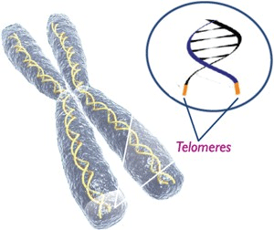
This observation combined with their 2010 research, shows Zscan4 is involved in elongating telomeres. Telomeres are the caps at the end of each strand of DNA which protect chromosomes. Researchers speculate longer cell cycles are triggered by Zscan4 to lengthen telomeres.
They also found that once cells expressed Zscan4, the next generation of cell cycles were shorter. This supports their idea that cell cycles slow down to allow for the recovery of telomeres — then speed up again when telomere repair is complete.
In a final test, researchers engineered stem cells to be deficient in Zscan4. They watched as deficient cells failed to recover from longer cell cycles — suffering cell death and proving their hypothesis.
Surprised by their findings, the authors have a new appreciation for how Zscan4 and pluripotent cells maintain their ability to replicate. A question now remaining is the relationship between Zscan4 and telomerase, the enzyme critical to telomere repair.
Abstract Highlights
•At longer cell cycles, telomeres are shorter
•Zscan4 is activated when the cell cycles become long
•After the activation of Zscan4, the next cell cycle becomes short
•We propose Zscan4 is activated for telomere maintenance irrespective of pluripotency
Summary
ZSCAN4 is a DNA-binding protein that functions for telomere elongation and genomic stability. In vivo, it is specifically expressed at the two-cell stage during mouse development. In vitro, it is transiently expressed in mouse embryonic stem cells (ESCs), only in 5% of the population at one time. Here we attempted to elucidate when, under what circumstances, Zscan4 is activated in ESCs. Using live cell imaging, we monitored the activity of Zscan4 together with the pluripotency marker Rex1. The lengths of the cell cycles in ESCs were diverse. Longer cell cycles were accompanied by shorter telomeres and higher activation of Zscan4. Since activation of Zscan4 is involved in telomere elongation, we speculate that the extended cell cycles accompanied by Zscan4 activation reflect the time for telomere recovery. Rex1 and Zscan4 did not show any correlation. Taken together, we propose that Zscan4 is activated to recover shortened telomeres during extended cell cycles, irrespective of the pluripotent status.
This is an open access article under the CC BY license (http://creativecommons.org/licenses/by/4.0/).
Return to top of page
|
|
|
Mar 25, 2016 Fetal Timeline Maternal Timeline News News Archive

Image Credit:
Hitoshi Niwa Laboratory, RIKEN Center for Developmental Biology
|
|
|
|


