|
|
Single-cell embryos can be chimeras
Researchers have found errors in single cell embryos which can lead to entire sets of maternal and paternal chromosomes being unevenly distributed — making them chimeras.
A chimera is an embryo made up of cells from different zygotes — a zygote being the fusion of DNA from each sex cell (gamete). One from dad and one from mom, in the typical embryo, but with chimeras, well, there are no rules. Chimeras can result in male and female organs in one body, two blood types in one child, or numerous other more subtle variations - but birth defects that need to be managed.
Differences of chimeras are often due to mutations occuring during ordinary cell division. Normally, chimerism is not visible by casual inspection, but detected when trying to prove parentage or understand the origin of a birth defect.
Single-cell embryos typically have one set each of maternal and paternal chromosomes. As the embryo grows, daughter cells bud off with their own copy of each set, leaving a stem cell behind to keep replicating daughter cells and building tissues for the growing embryo.
Previous studies involving in vitro fertilized (IVF) human embryos have demonstrated that large fractions of embryos contain at least one cell with either whole or partial gains or losses in numbers of chromosomes. Chromosomal instability can lead to reduced fecundity of the parents, as well as birth defects in their children.
To investigate this phenomenon in more detail, researchers from The Catholic University of Leuven or The Katholieke Universiteit Leuve (KU Leuven) in Leuven, Holland, along with other collaborators, employed in vitro fertilization in cattle as an animal model system to study chromosomal changes in single embryonic cells.
The work is published in Genome Research.
"This is a novel fundamental insight into the origin of chimerism, a very rare condition in humans which can lead to birth defects."
Joris Vermeesh PhD, Group Leader, Laboratory for Cytogenetics and Genome Research, The Catholic University of Leuven, and senior author.
Applying a method they had previously developed, KU Leuven researchers examined number and parental origin of chromosomes in single cells from 23 cow embryos. Nearly three-fourths of the embryos contained at least one cell with either unexpected partial or whole chromosome abnomalities, similar to what is found in human in-vitro fertilized embryos.
Surprisingly, 39% of all embryos contained cells with abnormalities in entire sets of embryos. For example, cells with only one set of chromosomes, either maternal or paternal, or three sets (one maternal set plus two paternal sets, or vice versa).
Many of these embryos resulted from fertilization errors, such as two sperm fertilizing a single egg.
However, normally fertilized embryos also displayed these aberrant patterns. Researchers named the process of cell division leading to segregation of parental chromosomes as "heterogoneic," or of differential parental origin.
Finding that normal fertilization can result in embryos containing cells with different parental sets of chromosomes is new — and a new mechanism for chimerism. Chimeras were previously thought to occur only as the result of mistakes in fertilization. For example, fusion of multiple sperm to an egg (or eggs) that somehow continue on to form an entire organism.
"The presence of chimerism in human IVF embryos was never thought of as happening before this study. Knowing this might occur may improve approaches for embryo selection and ultimately [improve] the success of IVF pre-implantation genetic diagnosis."
Joris Vermeesh PhD
Abstract
Dramatic genome dynamics, such as chromosome instability, contribute to the remarkable genomic heterogeneity among the blastomeres comprising a single embryo during human preimplantation development. This heterogeneity, when compatible with life, manifests as constitutional mosaicism, chimerism, and mixoploidy in live-born individuals. Chimerism and mixoploidy are defined by the presence of cell lineages with different parental genomes or different ploidy states in a single individual, respectively. Our knowledge of their mechanistic origin results from indirect observations, often when the cell lineages have been subject to rigorous selective pressure during development. Here, we applied haplarithmisis to infer the haplotypes and the copy number of parental genomes in 116 single blastomeres comprising entire preimplantation bovine embryos (n = 23) following in vitro fertilization. We not only demonstrate that chromosome instability is conserved between bovine and human cleavage embryos, but we also discovered that zygotes can spontaneously segregate entire parental genomes into different cell lineages during the first post-zygotic cleavage division. Parental genome segregation was not exclusively triggered by abnormal fertilizations leading to triploid zygotes, but also normally fertilized zygotes can spontaneously segregate entire parental genomes into different cell lineages during cleavage of the zygote. We coin the term “heterogoneic division” to indicate the events leading to noncanonical zygotic cytokinesis, segregating the parental genomes into distinct cell lineages. Persistence of those cell lines during development is a likely cause of chimerism and mixoploidy in mammals.
Scientists from KU Leuven, Ghent University, University of Tartu, and Wellcome Trust Sanger Institute contributed to this study. The study was funded by the Agency for Innovation by Science and Technology, the Research Foundation Flanders, the European Union's Research and Innovation funding program and the University of Leuven.
About the article:
The manuscript will be published online ahead of print on 12 April 2016. Its full citation is as follows:
Destouni A, Zamani Esteki M, Catteeuw M, Tšuiko M, Dimitriadou E, Smits K, Kurg A, Van Soom A, Voet T, Vermeesch JR. 2016. Zygotes segregate entire parental genomes in distinct blastomere lineages causing cleavage stage chimerism and mixoploidy. Genome Res doi: 10.1101/gr.200527.115
The article is distributed exclusively by Cold Spring Harbor Laboratory Press for the first six months after the full-issue publication date (see http://genome.cshlp.org/site/misc/terms.xhtml). After six months, it is available under a Creative Commons License (Attribution-NonCommercial 4.0 International), as described at http://creativecommons.org/licenses/by-nc/4.0/.
About Genome Research:
Launched in 1995, Genome Research is an international, continuously published, peer-reviewed journal that focuses on research that provides novel insights into the genome biology of all organisms, including advances in genomic medicine. Among the topics considered by the journal are genome structure and function, comparative genomics, molecular evolution, genome-scale quantitative and population genetics, proteomics, epigenomics, and systems biology. The journal also features exciting gene discoveries and reports of cutting-edge computational biology and high-throughput methodologies.
About Cold Spring Harbor Laboratory Press:
Cold Spring Harbor Laboratory Press is an internationally renowned publisher of books, journals, and electronic media, located on Long Island, New York. Since 1933, it has furthered the advance and spread of scientific knowledge in all areas of genetics and molecular biology, including cancer biology, plant science, bioinformatics, and neurobiology. The Press is a division of Cold Spring Harbor Laboratory, an innovator in life science research and the education of scientists, students, and the public. For more information, visit our website at http://cshlpress.org/
Return to top of page
|
|
|
Apr 14, 2016 Fetal Timeline Maternal Timeline News News Archive
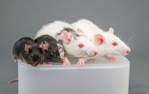
Injecting cells from one species into the embryo of another creates mixtures called chimeras.
Left to Right: Ordinary mouse, a mouse that’s partly rat, a rat that’s partly mouse, a white rat.
Image Credit: MIT Technology Review
|
|
|
|


