Welcome to The Visible Embryo
  o
Home-- -History-- -Bibliography- -Pregnancy Timeline- --Prescription Drugs in Pregnancy- -- Pregnancy Calculator- --Female Reproductive System- -Contact

Welcome to The Visible Embryo, a comprehensive educational resource on human development from conception to birth.

The Visible Embryo provides visual references for changes in fetal development throughout pregnancy and can be navigated via fetal development or maternal changes.

The National Institutes of Child Health and Human Development awarded Phase I and Phase II Small Business Innovative Research Grants to develop The Visible Embryo. Initally designed to evaluate the internet as a teaching tool for first year medical students, The Visible Embryo is linked to over 600 educational institutions and is viewed by more than one million visitors each month.

Today, The Visible Embryo is linked to over 600 educational institutions and is viewed by more than 1 million visitors each month. The field of early embryology has grown to include the identification of the stem cell as not only critical to organogenesis in the embryo, but equally critical to organ function and repair in the adult human. The identification and understanding of genetic malfunction, inflammatory responses, and the progression in chronic disease, begins with a grounding in primary cellular and systemic functions manifested in the study of the early embryo.

WHO International Clinical Trials Registry Platform


The World Health Organization (WHO) has created a new Web site to help researchers, doctors and
patients obtain reliable information on high-quality clinical trials. Now you can go to one website and search all registers to identify clinical trial research underway around the world!


Home

History

Bibliography

Pregnancy Timeline

Prescription Drug Effects on Pregnancy

Pregnancy Calculator

Female Reproductive System

Contact The Visible Embryo

News Alerts Archive

Disclaimer: The Visible Embryo web site is provided for your general information only. The information contained on this site should not be treated as a substitute for medical, legal or other professional advice. Neither is The Visible Embryo responsible or liable for the contents of any websites of third parties which are listed on this site.
Content protected under a Creative Commons License.

No dirivative works may be made or used for commercial purposes.


Pregnancy Timeline by SemestersLungs begin to produce surfactantImmune system beginningHead may position into pelvisFull TermPeriod of rapid brain growthWhite fat begins to be madeHead may position into pelvisWhite fat begins to be madeImmune system beginningBrain convolutions beginBrain convolutions beginFetal liver is producing blood cellsSensory brain waves begin to activateSensory brain waves begin to activateInner Ear Bones HardenBone marrow starts making blood cellsBone marrow starts making blood cellsBrown fat surrounds lymphatic systemFetal sexual organs visibleFinger and toe prints appearFinger and toe prints appearHeartbeat can be detectedHeartbeat can be detectedBasic Brain Structure in PlaceThe Appearance of SomitesFirst Detectable Brain WavesA Four Chambered HeartBeginning Cerebral HemispheresFemale Reproductive SystemEnd of Embryonic PeriodEnd of Embryonic PeriodFirst Thin Layer of Skin AppearsThird TrimesterSecond TrimesterFirst TrimesterFertilizationDevelopmental Timeline
CLICK ON weeks 0 - 40 and follow along every 2 weeks of fetal development
Google Search artcles published since 2007


Home | Pregnancy Timeline | News Alerts |News Archive Mar 12, 2015

An aberration in the CTCF folding of the Hox 6 gene, results in extra ribs in this mouse.


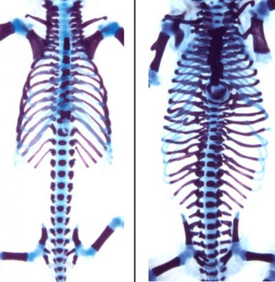
DNA-binding protein critical to normal embryo

Scientists have found that CTCF, a DNA-binding protein, is essential in the body plan of a developing embryo.

Scientists at NYU Langone Medical Center and New York University, studied mouse brain cells and how they manage animal movements. Their research led to important details on how Hox genes keep cells in position — as well as in the right order front to back.

The work was published in Science, February 27, 2015.


Hox genes are arranged in organized clusters on an animal genome, but only a subset of Hox genes are active at any given time in a cell.

Researchers observed that a precise "memory" from mother cell to daughter cell of active and inactive Hox genes is fundamental to a normal body plan. If a failure occurs in that "memory" system, a body part could be produced in the wrong anatomical position.


"Previous research has shown that CTCF acts as a key insulating barrier preventing mistakes in cells as they multiply and differentiate," says Varun Narendra, the study's lead author, and a fifth-year graduate PhD student in developmental biology at NYU Langone and the Howard Hughes Medical Institute. "Now we have shown that correct positioning also depends on CTCF."


"The findings provide new insight into how cells faithfully transmit organizational information as embryos develop. When cell development goes awry, abnormal development and diseases such as cancer can occur. Information from this study could help in therapies to address developmental missteps in Hox genes and their regulators."

Danny Reinberg PhD, professor of biochemistry and molecular pharmacology at NYU Langone, Howard Hughes Medical Institute investigator and senior study investigator.

CTCF is a DNA-binding protein, which marks "insulator" regions on the DNA of animals. These regions act as boundaries affecting how cells package DNA. In specific regions, CTCF binding ensures segments of the genome are packaged to become active, while not interfering with neighboring segments that should not become active in any daughter cells generated. Using mouse embryo stem cells that generate motor neurons, scientists identified how CTCF isolates Hox genes in this way.


"We found that the purpose of CTCF is to divide the Hox cluster into segments, allowing a cluster to fold into strict domains that are either active or inactive on either side of CTCF."

Varun Narendra, lead author, graduate PhD student.


To demonstrate that CTCF binding is needed for correct Hox gene activation, researchers removed sites on the genome where CTCF would normally bind, and discovered that without CTCF binding, the Hox cluster would not fold properly. As a result, motor neurons activated the wrong set of Hox genes.

"By altering the folding pattern of the Hox cluster, we altered the motor neurons' understanding of their anatomical position. In doing so, we also altered their ability to send nerve signals to the appropriate muscle targets," said Esteban Mazzoni, PhD, a study co-investigator and assistant professor of biology and New York University.


Because the precise activation of Hox genes is essential for a cell's fate, Mazzoni says: "the research should prove extremely useful in developing novel embryonic stem cell-based therapies."


Abstract
Polycomb and Trithorax group proteins encode the epigenetic memory of cellular positional identity by establishing inheritable domains of repressive and active chromatin within the Hox clusters. Here we demonstrate that the CCCTC-binding factor (CTCF) functions to insulate these adjacent yet antagonistic chromatin domains during embryonic stem cell differentiation into cervical motor neurons. Deletion of CTCF binding sites within the Hox clusters results in the expansion of active chromatin into the repressive domain. CTCF functions as an insulator by organizing Hox clusters into spatially disjoint domains. Ablation of CTCF binding disrupts topological boundaries such that caudal Hox genes leave the repressed domain and become subject to transcriptional activation. Hence, CTCF is required to insulate facultative heterochromatin from impinging euchromatin to produce discrete positional identities.

Other contributors to the study, all from NYU Langone's Department of Biochemistry and Molecular Pharmacology, are Pedro P. Rocha, PhD; Disi An; Ramya Raviram, and Jane A. Skok, PhD.

Funding support for this study was provided by The Howard Hughes Medical Institute, National Institute of Health grants (R37-37120, RM-64844, T32 GM08652, GM112192, and R01 HD079682) and the P.A.L.S. Foundation.

Return to top of page